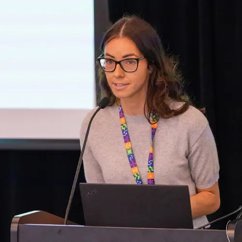
Portrait de Julien Cohen-Adad

Julien Cohen-Adad

Membre académique associé
Professeur agrégé, Polytechnique Montréal, Département de génie électrique
Professeur asssocié, Université de Montréal, Département de neurosciences
Sujets de recherche
Apprentissage automatique médical

Biographie

Julien Cohen-Adad est professeur à Polytechnique Montréal et directeur associé de l'Unité de neuro-imagerie fonctionnelle de l'Université de Montréal. Il est également titulaire de la Chaire de recherche du Canada en imagerie par résonance magnétique quantitative. Ses recherches portent sur l'avancement des méthodes de neuro-imagerie avec l'aide de l'IA. Voici quelques exemples de ses projets :

- Formation multimodale pour les tâches d'imagerie médicale (segmentation des pathologies, diagnostic, etc.);

- Ajout d'un a priori issu de la physique de l'IRM pour améliorer la généralisation des modèles;

- Incorporation de mesures d'incertitude pour traiter la variabilité interévaluateurs;

- Stratégies d'apprentissage continu lorsque le partage des données est restreint;

- Introduction des méthodes d'IA dans la routine de la radiologie clinique par l’intermédiaire de solutions logicielles conviviales.

Le professeur Cohen-Adad dirige également de nombreux projets de logiciels libres qui profitent à la communauté scientifique et clinique. Plus de détails sur https://neuro.polymtl.ca/software.html.

En résumé, Julien aime : l'IRM avec des aimants puissants, la neuro-imagerie, la programmation et la science ouverte!

Étudiants actuels

Collaborateur·rice alumni - Polytechnique
Co-superviseur⋅e :
Stagiaire de recherche - Polytechnique
Doctorat - Polytechnique
Co-superviseur⋅e :
Doctorat - Polytechnique
Maîtrise recherche - Polytechnique
Doctorat - Polytechnique
Collaborateur·rice de recherche
Maîtrise recherche - UdeM
Maîtrise recherche - Polytechnique
Postdoctorat - Polytechnique

Publications

Longitudinal reproducibility of brain and spinal cord quantitative MRI biomarkers
Mathieu Boudreau
Agah Karakuzu
Arnaud Boré
Basile Pinsard
Kiril Zelenkovski
Eva Alonso‐Ortiz
Julie Boyle
Quantitative MRI (qMRI) promises better specificity, accuracy, repeatability, and reproducibility relative to its clinically-used qualitativ… (voir plus)e MRI counterpart. Longitudinal reproducibility is particularly important in qMRI. The goal is to reliably quantify tissue properties that may be assessed in longitudinal clinical studies throughout disease progression or during treatment. In this work, we present the initial data release of the quantitative MRI portion of the Courtois project on neural modelling (CNeuroMod), where the brain and cervical spinal cord of six participants were scanned at regular intervals over the course of several years. This first release includes three years of data collection and up to ten sessions per participant using quantitative MRI imaging protocols (T1, magnetization transfer (MTR, MTsat), and diffusion). In the brain, T1MP2RAGE, FA, MD, and RD all exhibited high longitudinal reproducibility (intraclass correlation coefficient— ICC ≃ 1 and within-subject coefficient of variations— wCV 1%). The spinal cord cross-sectional area (CSA) computed using T2w images and T1MTsat exhibited the best longitudinal reproducibility (ICC ≃ 1 and 0.7 respectively, and wCV 2.4% and 6.9%). Results from this work show the level of longitudinal reproducibility that can be expected from qMRI protocols in the brain and spinal cord in the absence of hardware and software upgrades, and could help in the design of future longitudinal clinical studies.
Body size and intracranial volume interact with the structure of the central nervous system: A multi-center in vivo neuroimaging study
René Labounek
Monica T. Bondy
Amy L. Paulson
Sandrine Bédard
Mihael Abramovic
Eva Alonso‐Ortiz
Nicole Atcheson
Laura R. Barlow
Robert L. Barry
Markus Barth
Marco Battiston
Christian Büchel
Matthew D. Budde
Virginie Callot
Anna Combes
Benjamin De Leener
Maxime Descoteaux
Paulo Loureiro de Sousa
Marek Dostál
Julien Doyon … (voir 74 de plus)
Adam Dvorak
Falk Eippert
Karla R. Epperson
Kevin S. Epperson
Patrick Freund
Jürgen Finsterbusch
Alexandru Foias
Michela Fratini
Issei Fukunaga
Claudia A. M. Gandini Wheeler-Kingshott
Giancarlo Germani
Guillaume Gilbert
Federico Giove
Francesco Grussu
Akifumi Hagiwara
Pierre-Gilles Henry
Tomáš Horák
Masaaki Hori
James Joers
Kouhei Kamiya
Haleh Karbasforoushan
Miloš Keřkovský
Ali Khatibi
Joo-won Kim
Nawal Kinany
Hagen H. Kitzler
Shannon Kolind
Yazhuo Kong
Petr Kudlička
Paul Kuntke
Nyoman D. Kurniawan
Slawomir Kusmia
Maria Marcella Lagana
Cornelia Laule
Christine S. W. Law
Csw Law
Tobias Leutritz
Yaou Liu
Sara Llufriu
Sean Mackey
Allan R. Martin
Eloy Martinez-Heras
Loan Mattera
Kristin P. O’Grady
Nico Papinutto
Daniel Papp
Deborah Pareto
Todd B. Parrish
Anna Pichiecchio
Ferran Prados
Àlex Rovira
Marc J. Ruitenberg
Rebecca S. Samson
Giovanni Savini
Maryam Seif
Alan C. Seifert
Alex K. Smith
Seth Aaron Smith
Zachary A. Smith
Elisabeth Solana
Yuichi Suzuki
George Tackley
Alexandra Tinnermann
Dimitri Van De Ville
Marios C. Yiannakas
Kenneth A. Weber
Nikolaus Weiskopf
Richard G. Wise
Patrik O. Wyss
Junqian Xu
Christophe Lenglet
Igor Nestrašil
Longitudinal reproducibility of brain and spinal cord quantitative MRI biomarkers
Mathieu Boudreau
Agah Karakuzu
Arnaud Boré
Basile Pinsard
Kiril Zelenkovski
Eva Alonso‐Ortiz
Julie Boyle
Abstract Quantitative MRI (qMRI) promises better specificity, accuracy, repeatability, and reproducibility relative to its clinically-used q… (voir plus)ualitative MRI counterpart. Longitudinal reproducibility is particularly important in qMRI. The goal is to reliably quantify tissue properties that may be assessed in longitudinal clinical studies throughout disease progression or during treatment. In this work, we present the initial data release of the quantitative MRI portion of the Courtois project on neural modelling (CNeuroMod), where the brain and cervical spinal cord of six participants were scanned at regular intervals over the course of several years. This first release includes 3 years of data collection and up to 10 sessions per participant using quantitative MRI imaging protocols (T1, magnetization transfer (MTR, MTsat), and diffusion). In the brain, T1MP2RAGE, fractional anisotropy (FA), mean diffusivity (MD), and radial diffusivity (RD) all exhibited high longitudinal reproducibility (intraclass correlation coefficient – ICC ≃ 1 and within-subject coefficient of variations – wCV 1%). The spinal cord cross-sectional area (CSA) computed using T2w images and T1MTsat exhibited the best longitudinal reproducibility (ICC ≃ 1 and 0.7 respectively, and wCV 2.4% and 6.9%). Results from this work show the level of longitudinal reproducibility that can be expected from qMRI protocols in the brain and spinal cord in the absence of hardware and software upgrades, and could help in the design of future longitudinal clinical studies.
Spinal cord demyelination predicts neurological deterioration in patients with mild degenerative cervical myelopathy
Abdul Al-Shawwa
Michael Craig
David Anderson
Steven Casha
W. Bradley Jacobs
Nathan Evaniew
Saswati Tripathy
Jacques Bouchard
Peter Lewkonia
Fred Nicholls
Alex Soroceanu
Ganesh Swamy
Kenneth C. Thomas
Stephan duPlessis
Michael M.H. Yang
Nicholas Dea
Jefferson R. Wilson
David W. Cadotte
Towards contrast-agnostic soft segmentation of the spinal cord
Sandrine Bédard
Enamundram Naga Karthik
Charidimos Tsagkas
Emanuele Pravatà
Cristina Granziera
Andrew C. Smith
Kenneth Arnold Weber
Spinal cord segmentation is clinically relevant and is notably used to compute spinal cord cross-sectional area (CSA) for the diagnosis and … (voir plus)monitoring of cord compression or neurodegenerative diseases such as multiple sclerosis. While several semi and automatic methods exist, one key limitation remains: the segmentation depends on the MRI contrast, resulting in different CSA across contrasts. This is partly due to the varying appearance of the boundary between the spinal cord and the cerebrospinal fluid that depends on the sequence and acquisition parameters. This contrast-sensitive CSA adds variability in multi-center studies where protocols can vary, reducing the sensitivity to detect subtle atrophies. Moreover, existing methods enhance the CSA variability by training one model per contrast, while also producing binary masks that do not account for partial volume effects. In this work, we present a deep learning-based method that produces soft segmentations of the spinal cord. Using the Spine Generic Public Database of healthy participants (
TransCeption: Enhancing medical image segmentation with an inception-like transformer design for efficient feature fusion
Reza Azad
Yiwei Jia
Ehsan Khodapanah Aghdam
Dorit Merhof
SCIseg: Automatic Segmentation of Intramedullary Lesions in Spinal Cord Injury on T2-weighted MRI Scans.
Enamundram Naga Karthik
Andrew C. Smith
Dario Pfyffer
Simon Schading-Sassenhausen
Lynn Farner
KA Weber
Patrick Freund
"Just Accepted" papers have undergone full peer review and have been accepted for publication in Radiology: Artificial Intelligence. This ar… (voir plus)ticle will undergo copyediting, layout, and proof review before it is published in its final version. Please note that during production of the final copyedited article, errors may be discovered which could affect the content. Purpose To develop a deep learning tool for the automatic segmentation of the spinal cord and intramedullary lesions in spinal cord injury (SCI) on T2-weighted MRI scans. Materials and Methods This retrospective study included MRI data acquired between July 2002 and February 2023 from 191 patients with SCI (mean age, 48.1 years ± 17.9 [SD]; 142 males). The data consisted of T2-weighted MRI acquired using different scanner manufacturers with various image resolutions (isotropic and anisotropic) and orientations (axial and sagittal). Patients had different lesion etiologies (traumatic, ischemic, and hemorrhagic) and lesion locations across the cervical, thoracic and lumbar spine. A deep learning model, SCIseg, was trained in a three-phase process involving active learning for the automatic segmentation of intramedullary SCI lesions and the spinal cord. The segmentations from the proposed model were visually and quantitatively compared with those from three other open-source methods (PropSeg, DeepSeg and contrast-agnostic, all part of the Spinal Cord Toolbox). Wilcoxon signed-rank test was used to compare quantitative MRI biomarkers of SCI (lesion volume, lesion length, and maximal axial damage ratio) derived from the manual reference standard lesion masks and biomarkers obtained automatically with SCIseg segmentations. Results SCIseg achieved a Dice score of 0.92 ± 0.07 (mean ± SD) and 0.61 ± 0.27 for spinal cord and SCI lesion segmentation, respectively. There was no evidence of a difference between lesion length (P = .42) and maximal axial damage ratio (P = .16) computed from manually annotated lesions and the lesion segmentations obtained using SCIseg. Conclusion SCIseg accurately segmented intramedullary lesions on a diverse dataset of T2-weighted MRI scans and extracted relevant lesion biomarkers (namely, lesion volume, lesion length, and maximal axial damage ratio). SCIseg is open-source and accessible through the Spinal Cord Toolbox (v6.2 and above). Published under a CC BY 4.0 license.
SCIseg: Automatic Segmentation of Intramedullary Lesions in Spinal Cord Injury on T2-weighted MRI Scans.
Enamundram Naga Karthik
Andrew C. Smith
Dario Pfyffer
Simon Schading-Sassenhausen
Lynn Farner
KA Weber
Kenneth A. Weber
Patrick Freund
"Just Accepted" papers have undergone full peer review and have been accepted for publication in Radiology: Artificial Intelligence. This ar… (voir plus)ticle will undergo copyediting, layout, and proof review before it is published in its final version. Please note that during production of the final copyedited article, errors may be discovered which could affect the content. Purpose To develop a deep learning tool for the automatic segmentation of the spinal cord and intramedullary lesions in spinal cord injury (SCI) on T2-weighted MRI scans. Materials and Methods This retrospective study included MRI data acquired between July 2002 and February 2023 from 191 patients with SCI (mean age, 48.1 years ± 17.9 [SD]; 142 males). The data consisted of T2-weighted MRI acquired using different scanner manufacturers with various image resolutions (isotropic and anisotropic) and orientations (axial and sagittal). Patients had different lesion etiologies (traumatic, ischemic, and hemorrhagic) and lesion locations across the cervical, thoracic and lumbar spine. A deep learning model, SCIseg, was trained in a three-phase process involving active learning for the automatic segmentation of intramedullary SCI lesions and the spinal cord. The segmentations from the proposed model were visually and quantitatively compared with those from three other open-source methods (PropSeg, DeepSeg and contrast-agnostic, all part of the Spinal Cord Toolbox). Wilcoxon signed-rank test was used to compare quantitative MRI biomarkers of SCI (lesion volume, lesion length, and maximal axial damage ratio) derived from the manual reference standard lesion masks and biomarkers obtained automatically with SCIseg segmentations. Results SCIseg achieved a Dice score of 0.92 ± 0.07 (mean ± SD) and 0.61 ± 0.27 for spinal cord and SCI lesion segmentation, respectively. There was no evidence of a difference between lesion length (P = .42) and maximal axial damage ratio (P = .16) computed from manually annotated lesions and the lesion segmentations obtained using SCIseg. Conclusion SCIseg accurately segmented intramedullary lesions on a diverse dataset of T2-weighted MRI scans and extracted relevant lesion biomarkers (namely, lesion volume, lesion length, and maximal axial damage ratio). SCIseg is open-source and accessible through the Spinal Cord Toolbox (v6.2 and above). Published under a CC BY 4.0 license.
Spinal cord evaluation in multiple sclerosis: clinical and radiological associations, present and future
B Mark Keegan
Martina Absinta
Eoin P Flanagan
Roland G Henry
Eric C Klawiter
Shannon Kolind
Stephen Krieger
Cornelia Laule
John A Lincoln
Steven Messina
Jiwon Oh
Nico Papinutto
Seth Aaron Smith
Anthony Traboulsee
Abstract Spinal cord disease is important in most people with multiple sclerosis, but assessment remains less emphasized in patient care, ba… (voir plus)sic and clinical research and therapeutic trials. The North American Imaging in Multiple Sclerosis Spinal Cord Interest Group was formed to determine and present the contemporary landscape of multiple sclerosis spinal cord evaluation, further existing and advanced spinal cord imaging techniques, and foster collaborative work. Important themes arose: (i) multiple sclerosis spinal cord lesions (differential diagnosis, association with clinical course); (ii) spinal cord radiological–pathological associations; (iii) ‘critical’ spinal cord lesions; (iv) multiple sclerosis topographical model; (v) spinal cord atrophy; and (vi) automated and special imaging techniques. Distinguishing multiple sclerosis from other myelopathic aetiology is increasingly refined by imaging and serological studies. Post-mortem spinal cord findings and MRI pathological correlative studies demonstrate MRI’s high sensitivity in detecting microstructural demyelination and axonal loss. Spinal leptomeninges include immune inflammatory infiltrates, some in B-cell lymphoid-like structures. ‘Critical’ demyelinating lesions along spinal cord corticospinal tracts are anatomically consistent with and may be disproportionately associated with motor progression. Multiple sclerosis topographical model implicates the spinal cord as an area where threshold impairment associates with multiple sclerosis disability. Progressive spinal cord atrophy and ‘silent’ multiple sclerosis progression may be emerging as an important multiple sclerosis prognostic biomarker. Manual atrophy assessment is complicated by rater bias, while automation (e.g. Spinal Cord Toolbox), and artificial intelligence may reduce this. Collaborative research by the North American Imaging in Multiple Sclerosis and similar groups with experts combining distinct strengths is key to advancing assessment and treatment of people with multiple sclerosis spinal cord disease.
Spiral volumetric optoacoustic tomography of reduced oxygen saturation in the spinal cord of M83 mouse model of Parkinson’s disease
Benjamin F. Combes
Sandeep Kumar Kalva
Pierre-Louis Benveniste
Agathe Tournant
Man Hoi Law
Joshua Newton
Maik Krüger
Rebecca Z Weber
Inês Dias
Daniela Noain
Xose Luis Dean-Ben
Uwe Konietzko
Christian R. Baumann
Per-Göran Gillberg
Christoph Hock
Roger M. Nitsch
Daniel Razansky
Ruiqing Ni