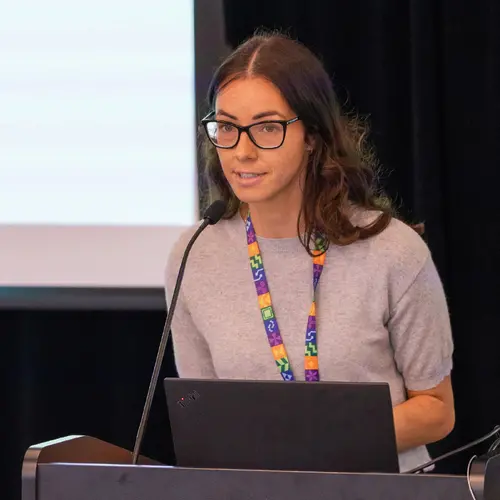
Portrait of Julien Cohen-Adad

Julien Cohen-Adad

Associate Academic Member
Associate Professor, Polytechnique Montréal, Electrical Engineering Department
Adjunct Professor, Université de Montréal, Department of Neuroscience
Research Topics
Medical Machine Learning

Biography

Julien Cohen-Adad is a professor at Polytechnique Montréal and the associate director of the Neuroimaging Functional Unit at Université de Montréal. He is also the Canada Research Chair in Quantitative Magnetic Resonance Imaging.

His research focuses on advancing neuroimaging methods with the help of AI. Some examples of projects are:

- Multi-modal training for medical imaging tasks (segmentation of pathologies, diagnosis, etc.)

- Adding prior from MRI physics to improve model generalization

- Incorporating uncertainty measures to deal with inter-rater variability

- Continuous learning strategies when data sharing is restricted

- Bringing AI methods into clinical radiology routine via user-friendly software solutions

Cohen-Adad also leads multiple open-source software projects that are benefiting the research and clinical community (see neuro.polymtl.ca/software.html). In short, he loves MRI with strong magnets, neuroimaging, programming and open science!

Current Students

Master's Research - Polytechnique Montréal
Co-supervisor :
PhD - Polytechnique Montréal
Co-supervisor :
PhD - Polytechnique Montréal
Master's Research - Polytechnique Montréal
PhD - Polytechnique Montréal
Collaborating researcher
Research Intern - Polytechnique Montréal
Master's Research - Université de Montréal
Master's Research - Polytechnique Montréal
Postdoctorate - Polytechnique Montréal

Publications

Myelin basic protein mRNA levels affect myelin sheath dimensions, architecture, plasticity, and density of resident glial cells
Hooman Bagheri
Hana Friedman
Amanda Hadwen
Celia Jarweh
Ellis Cooper
Lawrence Oprea
Claire Guerrier
Anmar Khadra
Armand Collin
Amanda Young
Gerardo Mendez Victoriano
Matthew Swire
Andrew Jarjour
Marie E. Bechler
Rachel S. Pryce
Pierre Chaurand
Lise Cougnaud
Dajana Vuckovic
Elliott Wilion … (see 11 more)
Owen Greene
Akiko Nishiyama
Anouk Benmamar‐Badel
Trevor Owens
Vladimir Grouza
Marius Tuznik
Hanwen Liu
David A. Rudko
Jinyi Zhang
Katherine A. Siminovitch
Alan C. Peterson
Abstract Myelin Basic Protein (MBP) is essential for both elaboration and maintenance of CNS myelin, and its reduced accumulation results in… (see more) hypomyelination. How different Mbp mRNA levels affect myelin dimensions across the lifespan and how resident glial cells may respond to such changes are unknown. Here, to investigate these questions, we used enhancer‐edited mouse lines that accumulate Mbp mRNA levels ranging from 8% to 160% of wild type. In young mice, reduced Mbp mRNA levels resulted in corresponding decreases in Mbp protein accumulation and myelin sheath thickness, confirming the previously demonstrated rate‐limiting role of Mbp transcription in the control of initial myelin synthesis. However, despite maintaining lower line specific Mbp mRNA levels into old age, both MBP protein levels and myelin thickness improved or fully normalized at rates defined by the relative Mbp mRNA level. Sheath length, in contrast, was affected only when mRNA levels were very low, demonstrating that sheath thickness and length are not equally coupled to Mbp mRNA level. Striking abnormalities in sheath structure also emerged with reduced mRNA levels. Unexpectedly, an increase in the density of all glial cell types arose in response to reduced Mbp mRNA levels. This investigation extends understanding of the role MBP plays in myelin sheath elaboration, architecture, and plasticity across the mouse lifespan and illuminates a novel axis of glial cell crosstalk.
Automatic Segmentation of the Spinal Cord Nerve Rootlets
Jan Valošek
Theo Mathieu
Raphaëlle Schlienger
Olivia S. Kowalczyk
Precise identification of spinal nerve rootlets is relevant to delineate spinal levels for the study of functional activity in the spinal co… (see more)rd. The goal of this study was to develop an automatic method for the semantic segmentation of spinal nerve rootlets from T2-weighted magnetic resonance imaging (MRI) scans. Images from two open-access MRI datasets were used to train a 3D multi-class convolutional neural network using an active learning approach to segment C2-C8 dorsal nerve rootlets. Each output class corresponds to a spinal level. The method was tested on 3T T2-weighted images from datasets unseen during training to assess inter-site, inter-session, and inter-resolution variability. The test Dice score was 0.67 +- 0.16 (mean +- standard deviation across rootlets levels), suggesting a good performance. The method also demonstrated low inter-vendor and inter-site variability (coefficient of variation= 1.41 %), as well as low inter-session variability (coefficient of variation= 1.30 %) indicating stable predictions across different MRI
Open Source in Lab Management
This document explores the advantages of integrating open source software and practices in managing a scientific lab, emphasizing reproducib… (see more)ility and the avoidance of pitfalls. It details practical applications from website management using GitHub Pages to organizing datasets in compliance with BIDS standards, highlights the importance of continuous testing for data integrity, IT management through Ansible for efficient system configuration, open source software development. The broader goal is to promote transparent, reproducible science by adopting open source tools. This approach not only saves time but exposes students to best practices, enhancing the transparency and reproducibility of scientific research.
Calibration-free parallel transmission of the cervical, thoracic, and lumbar spinal cord at 7T.
Christoph S. Aigner
Manuel F. Sánchez Alarcon
Alexandre D'Astous
Eva Alonso‐Ortiz
Sebastian Schmitter
PURPOSE To address the limitations of spinal cord imaging at ultra-high field (UHF) due to time-consuming parallel transmit (pTx) adjustment… (see more)s. This study introduces calibration-free offline computed universal shim modes that can be applied seamlessly for different pTx RF coils and spinal cord target regions, substantially enhancing spinal cord imaging efficiency at UHF. METHODS A library of channel-wise relative B 1 +
Calibration‐free parallel transmission of the cervical, thoracic, and lumbar spinal cord at <scp>7T</scp>
Christoph S. Aigner
Manuel F. Sánchez Alarcon
Alexandre D'Astous
Eva Alonso‐Ortiz
Sebastian Schmitter
Repeat it without me: Crowdsourcing the T1 mapping common ground via the ISMRM reproducibility challenge.
Mathieu Boudreau
Agah Karakuzu
Ecem Bozkurt
Madeline Carr
Marco Castellaro
Luis Concha
Mariya Doneva
Seraina A. Dual
Alex Ensworth
Alexandru Foias
Véronique Fortier
Refaat E. Gabr
Guillaume Gilbert
Carri K. Glide‐Hurst
Matthew Grech‐Sollars
Siyuan Hu
Oscar Jalnefjord
Jorge Jovicich
Kübra Keskin … (see 22 more)
Peter Koken
Anastasia Kolokotronis
Simran Kukran
Nam G. Lee
Ives R. Levesque
Bochao Li
Dan Ma
Burkhard Mädler
Nyasha G. Maforo
Jamie Near
Erick Pasaye
Alonso Ramirez‐Manzanares
Ben Statton
Christian Stehning
Stefano Tambalo
Ye Tian
Chenyang Wang
Kilian Weiss
Niloufar Zakariaei
Shuo Zhang
Ziwei Zhao
Nikola Stikov
PURPOSE T1 mapping is a widely used quantitative MRI technique, but its tissue-specific values remain inconsistent across protocols, sites, … (see more)and vendors. The ISMRM Reproducible Research and Quantitative MR study groups jointly launched a challenge to assess the reproducibility of a well-established inversion-recovery T1 mapping technique, using acquisition details from a seminal T1 mapping paper on a standardized phantom and in human brains. METHODS The challenge used the acquisition protocol from Barral et al. (2010). Researchers collected T1 mapping data on the ISMRM/NIST phantom and/or in human brains. Data submission, pipeline development, and analysis were conducted using open-source platforms. Intersubmission and intrasubmission comparisons were performed. RESULTS Eighteen submissions (39 phantom and 56 human datasets) on scanners by three MRI vendors were collected at 3 T (except one, at 0.35 T). The mean coefficient of variation was 6.1% for intersubmission phantom measurements, and 2.9% for intrasubmission measurements. For humans, the intersubmission/intrasubmission coefficient of variation was 5.9/3.2% in the genu and 16/6.9% in the cortex. An interactive dashboard for data visualization was also developed: https://rrsg2020.dashboards.neurolibre.org. CONCLUSION The T1 intersubmission variability was twice as high as the intrasubmission variability in both phantoms and human brains, indicating that the acquisition details in the original paper were insufficient to reproduce a quantitative MRI protocol. This study reports the inherent uncertainty in T1 measures across independent research groups, bringing us one step closer to a practical clinical baseline of T1 variations in vivo.
Repeat it without me: Crowdsourcing the T1 mapping common ground via the ISMRM reproducibility challenge
Mathieu Boudreau
Agah Karakuzu
Ecem Bozkurt
Madeline Carr
Marco Castellaro
Luis Concha
Mariya Doneva
Seraina A. Dual
Alex Ensworth
Alexandru Foias
Véronique Fortier
Refaat E. Gabr
Guillaume Gilbert
Carri K. Glide‐Hurst
Matthew Grech‐Sollars
Siyuan Hu
Oscar Jalnefjord
Jorge Jovicich
Kübra Keskin … (see 22 more)
Peter Koken
Anastasia Kolokotronis
Simran Kukran
Nam G. Lee
Ives R. Levesque
Bochao Li
Dan Ma
Burkhard Mädler
Nyasha G. Maforo
Jamie Near
Erick Pasaye
Alonso Ramirez‐Manzanares
Ben Statton
Christian Stehning
Stefano Tambalo
Ye Tian
Chenyang Wang
Kilian Weiss
Niloufar Zakariaei
Shuo Zhang
Ziwei Zhao
Nikola Stikov
T1 mapping is a widely used quantitative MRI technique, but its tissue‐specific values remain inconsistent across protocols, sites, and ve… (see more)ndors. The ISMRM Reproducible Research and Quantitative MR study groups jointly launched a challenge to assess the reproducibility of a well‐established inversion‐recovery T1 mapping technique, using acquisition details from a seminal T1 mapping paper on a standardized phantom and in human brains.
Quantifying neurodegeneration of the cervical cord and brain in degenerative cervical myelopathy: A multicentre study using quantitative <scp>magnetic resonance imaging</scp>
Patrick Freund
Viveka Boller
Tim M. Emmenegger
Muhammad Akbar
Markus Hupp
Nikolai Pfender
Claudia A. M. Gandini Wheeler-Kingshott
Michael G. Fehlings
Armin Curt
Maryam Seif
Quantifying neurodegeneration of the cervical cord and brain in degenerative cervical myelopathy: A multicentre study using quantitative magnetic resonance imaging
Patrick Freund
Viveka Boller
Tim M. Emmenegger
Muhammad Akbar
Markus Hupp
Nikolai Pfender
Claudia A. M. Gandini Wheeler-Kingshott
Michael G. Fehlings
Armin Curt
Maryam Seif
Simultaneous assessment of neurodegeneration in both the cervical cord and brain across multiple centres can enhance the effectiveness of cl… (see more)inical trials. Thus, this study aims to simultaneously assess microstructural changes in the cervical cord and brain above the stenosis in degenerative cervical myelopathy (DCM) using quantitative magnetic resonance imaging (MRI) in a multicentre study.