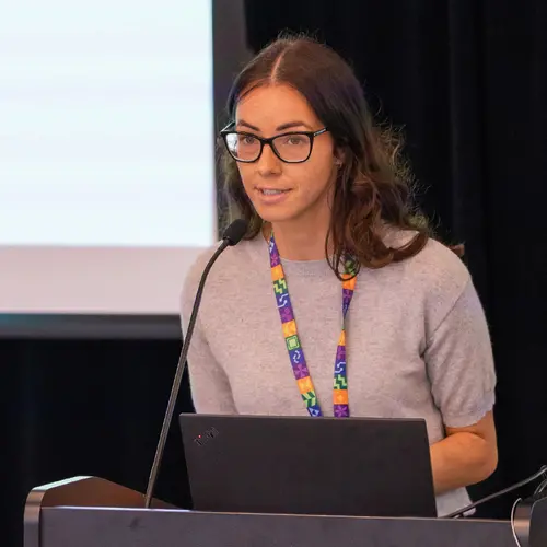
Portrait of Julien Cohen-Adad

Julien Cohen-Adad

Associate Academic Member
Associate Professor, Polytechnique Montréal, Electrical Engineering Department
Adjunct Professor, Université de Montréal, Department of Neuroscience
Research Topics
Medical Machine Learning

Biography

Julien Cohen-Adad is a professor at Polytechnique Montréal and the associate director of the Neuroimaging Functional Unit at Université de Montréal. He is also the Canada Research Chair in Quantitative Magnetic Resonance Imaging.

His research focuses on advancing neuroimaging methods with the help of AI. Some examples of projects are:

- Multi-modal training for medical imaging tasks (segmentation of pathologies, diagnosis, etc.)

- Adding prior from MRI physics to improve model generalization

- Incorporating uncertainty measures to deal with inter-rater variability

- Continuous learning strategies when data sharing is restricted

- Bringing AI methods into clinical radiology routine via user-friendly software solutions

Cohen-Adad also leads multiple open-source software projects that are benefiting the research and clinical community (see neuro.polymtl.ca/software.html). In short, he loves MRI with strong magnets, neuroimaging, programming and open science!

Current Students

Master's Research - Polytechnique Montréal
Co-supervisor :
PhD - Polytechnique Montréal
Co-supervisor :
PhD - Polytechnique Montréal
Master's Research - Polytechnique Montréal
PhD - Polytechnique Montréal
Collaborating researcher
Research Intern - Polytechnique Montréal
Master's Research - Université de Montréal
Master's Research - Polytechnique Montréal
Postdoctorate - Polytechnique Montréal

Publications

SCIseg: Automatic Segmentation of Intramedullary Lesions in Spinal Cord Injury on T2-weighted MRI Scans.
Enamundram Naga Karthik
Jan Valošek
Andrew C. Smith
Dario Pfyffer
Simon Schading-Sassenhausen
Lynn Farner
KA Weber
Kenneth A. Weber
Patrick Freund
"Just Accepted" papers have undergone full peer review and have been accepted for publication in Radiology: Artificial Intelligence. This ar… (see more)ticle will undergo copyediting, layout, and proof review before it is published in its final version. Please note that during production of the final copyedited article, errors may be discovered which could affect the content. Purpose To develop a deep learning tool for the automatic segmentation of the spinal cord and intramedullary lesions in spinal cord injury (SCI) on T2-weighted MRI scans. Materials and Methods This retrospective study included MRI data acquired between July 2002 and February 2023 from 191 patients with SCI (mean age, 48.1 years ± 17.9 [SD]; 142 males). The data consisted of T2-weighted MRI acquired using different scanner manufacturers with various image resolutions (isotropic and anisotropic) and orientations (axial and sagittal). Patients had different lesion etiologies (traumatic, ischemic, and hemorrhagic) and lesion locations across the cervical, thoracic and lumbar spine. A deep learning model, SCIseg, was trained in a three-phase process involving active learning for the automatic segmentation of intramedullary SCI lesions and the spinal cord. The segmentations from the proposed model were visually and quantitatively compared with those from three other open-source methods (PropSeg, DeepSeg and contrast-agnostic, all part of the Spinal Cord Toolbox). Wilcoxon signed-rank test was used to compare quantitative MRI biomarkers of SCI (lesion volume, lesion length, and maximal axial damage ratio) derived from the manual reference standard lesion masks and biomarkers obtained automatically with SCIseg segmentations. Results SCIseg achieved a Dice score of 0.92 ± 0.07 (mean ± SD) and 0.61 ± 0.27 for spinal cord and SCI lesion segmentation, respectively. There was no evidence of a difference between lesion length (P = .42) and maximal axial damage ratio (P = .16) computed from manually annotated lesions and the lesion segmentations obtained using SCIseg. Conclusion SCIseg accurately segmented intramedullary lesions on a diverse dataset of T2-weighted MRI scans and extracted relevant lesion biomarkers (namely, lesion volume, lesion length, and maximal axial damage ratio). SCIseg is open-source and accessible through the Spinal Cord Toolbox (v6.2 and above). Published under a CC BY 4.0 license.
Spinal cord evaluation in multiple sclerosis: clinical and radiological associations, present and future
B Mark Keegan
Martina Absinta
Eoin P Flanagan
Roland G Henry
Eric C Klawiter
Shannon Kolind
Stephen Krieger
Cornelia Laule
John A Lincoln
Steven Messina
Jiwon Oh
Nico Papinutto
Seth Aaron Smith
Anthony Traboulsee
Considerations and recommendations from the ISMRM Diffusion Study Group for preclinical diffusion MRI: Part 3 -- Ex vivo imaging: data processing, comparisons with microscopy, and tractography
Kurt G Schilling
Amy F. D. Howard
Francesco Grussu
Andrada Ianus
Brian Hansen
Rachel L. C. Barrett
Manisha Aggarwal
Stijn Michielse
Fatima Nasrallah
W. Syeda
Nian Wang
Jelle Veraart
Alard J. Roebroeck
Andrew F Bagdasarian
Cornelius Eichner
Farshid Sepehrband
Jan Zimmermann
L. Soustelle
Christien Bowman
Benjamin C. Tendler … (see 38 more)
A. Hertanu
Ben Jeurissen
Marleen Verhoye
Lucio Frydman
Y. Looij
David C. Hike
Jeff F. Dunn
Karla L. Miller
Bennett A. Landman
N. Shemesh
Adam Anderson
Emilie McKinnon
Shawna Farquharson
Flavio Dell’ Acqua
Carlo M. Pierpaoli
Ivana Drobnjak
Alexander Leemans
K. Harkins
Maxime Descoteaux
Duan Xu
Hao Huang
Mathieu D. Santin
Samuel C. Grant
Andre Obenaus
Gene S Kim
Dan Wu
D. Bihan
S. Blackband
Luisa Ciobanu
E. Fieremans
Ruiliang Bai
T. Leergaard
Jiangyang Zhang
T. Dyrby
G. A. Johnson
Matthew D. Budde
Ileana Ozana Jelescu
Spiral volumetric optoacoustic tomography of reduced oxygen saturation in the spinal cord of M83 mouse model of Parkinson's disease.
Benjamin F. Combes
Sandeep Kumar Kalva
Pierre-Louis Benveniste
Agathe Tournant
Man Hoi Law
Joshua Newton
Maik Krüger
Rebecca Z Weber
Inês Dias
Daniela Noain
Xose Luis Dean-Ben
Uwe Konietzko
Christian R. Baumann
Per-Göran Gillberg
Christoph Hock
Roger M. Nitsch
Daniel Razansky
Ruiqing Ni
<scp>RF</scp> shimming in the cervical spinal cord at <scp>7 T</scp>
Daniel Papp
Kyle M. Gilbert
Gaspard Cereza
Alexandre D'Astous
Nibardo Lopez‐Rios
Mathieu Boudreau
Marcus J. Couch
Pedram Yazdanbakhsh
Robert L. Barry
Eva Alonso‐Ortiz
SCIsegV2: A Universal Tool for Segmentation of Intramedullary Lesions in Spinal Cord Injury
Enamundram Naga Karthik
Jan Valošek
Lynn Farner
Dario Pfyffer
Simon Schading-Sassenhausen
A. Lebret
Gergely David
Andrew Smith
Kenneth A. Weber
Maryam Seif
Rhscir Network Imaging Group
Patrick Freund
Myelin basic protein mRNA levels affect myelin sheath dimensions, architecture, plasticity, and density of resident glial cells
Hooman Bagheri
Hana Friedman
Amanda Hadwen
Celia Jarweh
Ellis Cooper
Lawrence Oprea
Claire Guerrier
Anmar Khadra
Armand Collin
Amanda Young
Gerardo Mendez Victoriano
Matthew Swire
Andrew Jarjour
Marie E. Bechler
Rachel S. Pryce
Pierre Chaurand
Lise Cougnaud
Dajana Vuckovic
Elliott Wilion … (see 11 more)
Owen Greene
Akiko Nishiyama
Anouk Benmamar‐Badel
Trevor Owens
Vladimir Grouza
Marius Tuznik
Hanwen Liu
David A. Rudko
Jinyi Zhang
Katherine A. Siminovitch
Alan C. Peterson
Automatic Segmentation of the Spinal Cord Nerve Rootlets
Jan Valošek
Theo Mathieu
Raphaëlle Schlienger
Olivia S. Kowalczyk
Precise identification of spinal nerve rootlets is relevant to delineate spinal levels for the study of functional activity in the spinal co… (see more)rd. The goal of this study was to develop an automatic method for the semantic segmentation of spinal nerve rootlets from T2-weighted magnetic resonance imaging (MRI) scans. Images from two open-access MRI datasets were used to train a 3D multi-class convolutional neural network using an active learning approach to segment C2-C8 dorsal nerve rootlets. Each output class corresponds to a spinal level. The method was tested on 3T T2-weighted images from datasets unseen during training to assess inter-site, inter-session, and inter-resolution variability. The test Dice score was 0.67 +- 0.16 (mean +- standard deviation across rootlets levels), suggesting a good performance. The method also demonstrated low inter-vendor and inter-site variability (coefficient of variation= 1.41 %), as well as low inter-session variability (coefficient of variation= 1.30 %) indicating stable predictions across different MRI
Calibration‐free parallel transmission of the cervical, thoracic, and lumbar spinal cord at <scp>7T</scp>
Christoph S. Aigner
Manuel F. Sánchez Alarcon
Alexandre D'Astous
Eva Alonso‐Ortiz
Sebastian Schmitter
Repeat it without me: Crowdsourcing the T1 mapping common ground via the ISMRM reproducibility challenge.
Mathieu Boudreau
Agah Karakuzu
Ecem Bozkurt
Madeline Carr
Marco Castellaro
Luis Concha
Mariya Doneva
Seraina A. Dual
Alex Ensworth
Alexandru Foias
Véronique Fortier
Refaat E. Gabr
Guillaume Gilbert
Carri K. Glide‐Hurst
Matthew Grech‐Sollars
Siyuan Hu
Oscar Jalnefjord
Jorge Jovicich
Kübra Keskin … (see 22 more)
Peter Koken
Anastasia Kolokotronis
Simran Kukran
Nam G. Lee
Ives R. Levesque
Bochao Li
Dan Ma
Burkhard Mädler
Nyasha G. Maforo
Jamie Near
Erick Pasaye
Alonso Ramirez‐Manzanares
Ben Statton
Christian Stehning
Stefano Tambalo
Ye Tian
Chenyang Wang
Kilian Weiss
Niloufar Zakariaei
Shuo Zhang
Ziwei Zhao
Nikola Stikov
PURPOSE T1 mapping is a widely used quantitative MRI technique, but its tissue-specific values remain inconsistent across protocols, sites, … (see more)and vendors. The ISMRM Reproducible Research and Quantitative MR study groups jointly launched a challenge to assess the reproducibility of a well-established inversion-recovery T1 mapping technique, using acquisition details from a seminal T1 mapping paper on a standardized phantom and in human brains. METHODS The challenge used the acquisition protocol from Barral et al. (2010). Researchers collected T1 mapping data on the ISMRM/NIST phantom and/or in human brains. Data submission, pipeline development, and analysis were conducted using open-source platforms. Intersubmission and intrasubmission comparisons were performed. RESULTS Eighteen submissions (39 phantom and 56 human datasets) on scanners by three MRI vendors were collected at 3 T (except one, at 0.35 T). The mean coefficient of variation was 6.1% for intersubmission phantom measurements, and 2.9% for intrasubmission measurements. For humans, the intersubmission/intrasubmission coefficient of variation was 5.9/3.2% in the genu and 16/6.9% in the cortex. An interactive dashboard for data visualization was also developed: https://rrsg2020.dashboards.neurolibre.org. CONCLUSION The T1 intersubmission variability was twice as high as the intrasubmission variability in both phantoms and human brains, indicating that the acquisition details in the original paper were insufficient to reproduce a quantitative MRI protocol. This study reports the inherent uncertainty in T1 measures across independent research groups, bringing us one step closer to a practical clinical baseline of T1 variations in vivo.
Quantifying neurodegeneration of the cervical cord and brain in degenerative cervical myelopathy: A multicentre study using quantitative <scp>magnetic resonance imaging</scp>
Patrick Freund
Viveka Boller
Tim M. Emmenegger
Muhammad Akbar
Markus Hupp
Nikolai Pfender
Claudia A. M. Gandini Wheeler-Kingshott
Michael G. Fehlings
Armin Curt
Maryam Seif
Body size interacts with the structure of the central nervous system: A multi-center in vivo neuroimaging study
René Labounek
Monica T. Bondy
Amy L. Paulson
Sandrine Bédard
Mihael Abramovic
Eva Alonso‐Ortiz
Nicole Atcheson
Laura R. Barlow
Robert L. Barry
Markus Barth
Marco Battiston
Christian Büchel
Matthew D. Budde
Virginie Callot
Anna Combes
Benjamin De Leener
Maxime Descoteaux
Paulo Loureiro de Sousa
Marek Dostál
Julien Doyon … (see 74 more)
Adam Dvorak
Falk Eippert
Karla R. Epperson
Kevin S. Epperson
Patrick Freund
Jürgen Finsterbusch
Alexandru Foias
Michela Fratini
Issei Fukunaga
Claudia A. M. Gandini Wheeler-Kingshott
Giancarlo Germani
Guillaume Gilbert
Federico Giove
Francesco Grussu
Akifumi Hagiwara
Pierre-Gilles Henry
Tomáš Horák
Masaaki Hori
James M. Joers
Kouhei Kamiya
Haleh Karbasforoushan
Miloš Keřkovský
Ali Khatibi
Joo‐Won Kim
Nawal Kinany
Hagen H. Kitzler
Shannon Kolind
Yazhuo Kong
Petr Kudlička
Paul Kuntke
Nyoman D. Kurniawan
Slawomir Kusmia
Maria Marcella Lagana
Cornelia Laule
Christine S. W. Law
Csw Law
Tobias Leutritz
Yaou Liu
Sara Llufriu
Sean Mackey
Allan R. Martin
Eloy Martinez-Heras
Loan Mattera
Kristin P. O’Grady
Nico Papinutto
Daniel Papp
Deborah Pareto
Todd B. Parrish
Anna Pichiecchio
Ferran Prados
Àlex Rovira
Marc J. Ruitenberg
Rebecca S. Samson
Giovanni Savini
Maryam Seif
Alan C. Seifert
Alex K. Smith
Seth A. Smith
Zachary A. Smith
Elisabeth Solana
Yuichi Suzuki
George Tackley
Alexandra Tinnermann
Jan Valošek
Dimitri Van De Ville
Marios C. Yiannakas
Kenneth A. Weber
Nikolaus Weiskopf
Richard G. Wise
Patrik O. Wyss
Junqian Xu
Christophe Lenglet
Igor Nestrašil
Clinical research emphasizes the implementation of rigorous and reproducible study designs that rely on between-group matching or controllin… (see more)g for sources of biological variation such as subject’s sex and age. However, corrections for body size (i.e. height and weight) are mostly lacking in clinical neuroimaging designs. This study investigates the importance of body size parameters in their relationship with spinal cord (SC) and brain magnetic resonance imaging (MRI) metrics. Data were derived from a cosmopolitan population of 267 healthy human adults (age 30.1±6.6 years old, 125 females). We show that body height correlated strongly or moderately with brain gray matter (GM) volume, cortical GM volume, total cerebellar volume, brainstem volume, and cross-sectional area (CSA) of cervical SC white matter (CSA-WM; 0.44≤r≤0.62). In comparison, age correlated weakly with cortical GM volume, precentral GM volume, and cortical thickness (-0.21≥r≥-0.27). Body weight correlated weakly with magnetization transfer ratio in the SC WM, dorsal columns, and lateral corticospinal tracts (-0.20≥r≥-0.23). Body weight further correlated weakly with the mean diffusivity derived from diffusion tensor imaging (DTI) in SC WM (r=-0.20) and dorsal columns (-0.21), but only in males. CSA-WM correlated strongly or moderately with brain volumes (0.39≤r≤0.64), and weakly with precentral gyrus thickness and DTI-based fractional anisotropy in SC dorsal columns and SC lateral corticospinal tracts (-0.22≥r≥-0.25). Linear mixture of sex and age explained 26±10% of data variance in brain volumetry and SC CSA. The amount of explained variance increased at 33±11% when body height was added into the mixture model. Age itself explained only 2±2% of such variance. In conclusion, body size is a significant biological variable. Along with sex and age, body size should therefore be included as a mandatory variable in the design of clinical neuroimaging studies examining SC and brain structure.