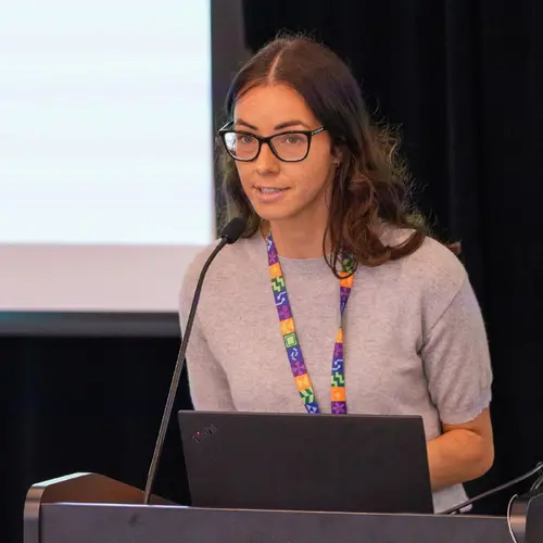
Portrait de Tal Arbel

Tal Arbel

Membre académique principal
Chaire en IA Canada-CIFAR
Professeure titulaire, McGill University, Département de génie électrique et informatique
Sujets de recherche
Apprentissage automatique médical
Apprentissage de représentations
Apprentissage profond
Causalité
Modèles génératifs
Modèles probabilistes
Vision par ordinateur

Biographie

Tal Arbel est professeure titulaire au Département de génie électrique et informatique de l’Université McGill, où elle dirige le groupe de vision probabiliste et le laboratoire d'imagerie médicale du Centre sur les machines intelligentes.

Elle est titulaire d'une chaire en IA Canada-CIFAR et membre associée de Mila – Institut québécois d’intelligence artificielle ainsi que du Centre de recherche sur le cancer Goodman. Les recherches de la professeure Arbel portent sur le développement de méthodes probabilistes d'apprentissage profond dans les domaines de la vision par ordinateur et de l’analyse d'imagerie médicale pour un large éventail d'applications dans le monde réel, avec un accent particulier sur les maladies neurologiques.

Elle a remporté le prix de la recherche Christophe Pierre 2019 de McGill Engineering et est Fellow à l'Académie canadienne d'ingénierie. Elle fait régulièrement partie de l'équipe organisatrice de grandes conférences internationales sur la vision par ordinateur et l'analyse d'imagerie médicale (par exemple celles de la Medical Image Computing and Computer-Assisted Intervention Society/MICCAI et de Medical Imaging with Deep Learning/MIDL, l’International Conference on Computer Vision/ICCV ou encore la Conference on Computer Vision and Pattern Recognition/CVPR). Elle est rédactrice en chef et cofondatrice de la revue Machine Learning for Biomedical Imaging (MELBA).


Étudiants actuels

Maîtrise recherche - McGill
Doctorat - McGill
Maîtrise recherche - McGill
Baccalauréat - McGill
Maîtrise recherche - McGill
Baccalauréat - McGill

Publications

Spatio-Temporal Conditional Diffusion Models for Forecasting Future Multiple Sclerosis Lesion Masks Conditioned on Treatments
Gian Mario Favero
Ge Ya Luo
Douglas Arnold
Spatio-Temporal Conditional Diffusion Models for Forecasting Future Multiple Sclerosis Lesion Masks Conditioned on Treatments
Gian Mario Favero
Ge Ya Luo
Douglas Arnold
Image-based personalized medicine has the potential to transform healthcare, particularly for diseases that exhibit heterogeneous progressio… (voir plus)n such as Multiple Sclerosis (MS). In this work, we introduce the first treatment-aware spatio-temporal diffusion model that is able to generate future masks demonstrating lesion evolution in MS. Our voxel-space approach incorporates multi-modal patient data, including MRI and treatment information, to forecast new and enlarging T2 (NET2) lesion masks at a future time point. Extensive experiments on a multi-centre dataset of 2131 patient 3D MRIs from randomized clinical trials for relapsing-remitting MS demonstrate that our generative model is able to accurately predict NET2 lesion masks for patients across six different treatments. Moreover, we demonstrate our model has the potential for real-world clinical applications through downstream tasks such as future lesion count and location estimation, binary lesion activity classification, and generating counterfactual future NET2 masks for several treatments with different efficacies. This work highlights the potential of causal, image-based generative models as powerful tools for advancing data-driven prognostics in MS.
AURA: A Multi-Modal Medical Agent for Understanding, Reasoning&Annotation
Recent advancements in Large Language Models (LLMs) have catalyzed a paradigm shift from static prediction systems to agentic AI agents capa… (voir plus)ble of reasoning, interacting with tools, and adapting to complex tasks. While LLM-based agentic systems have shown promise across many domains, their application to medical imaging remains in its infancy. In this work, we introduce AURA, the first visual linguistic explainability agent designed specifically for comprehensive analysis, explanation, and evaluation of medical images. By enabling dynamic interactions, contextual explanations, and hypothesis testing, AURA represents a significant advancement toward more transparent, adaptable, and clinically aligned AI systems. We highlight the promise of agentic AI in transforming medical image analysis from static predictions to interactive decision support. Leveraging Qwen-32B, an LLM-based architecture, AURA integrates a modular toolbox comprising: (i) a segmentation suite with phase grounding, pathology segmentation, and anatomy segmentation to localize clinically meaningful regions; (ii) a counterfactual image-generation module that supports reasoning through image-level explanations; and (iii) a set of evaluation tools including pixel-wise difference-map analysis, classification, and advanced state-of-the-art components to assess diagnostic relevance and visual interpretability.
Pixel Perfect MegaMed: A Megapixel-Scale Vision-Language Foundation Model for Generating High Resolution Medical Images
Medical image synthesis presents unique challenges due to the inherent complexity and high-resolution details required in clinical contexts.… (voir plus) Traditional generative architectures such as Generative Adversarial Networks (GANs) or Variational Auto Encoder (VAEs) have shown great promise for high-resolution image generation but struggle with preserving fine-grained details that are key for accurate diagnosis. To address this issue, we introduce Pixel Perfect MegaMed, the first vision-language foundation model to synthesize images at resolutions of 1024x1024. Our method deploys a multi-scale transformer architecture designed specifically for ultra-high resolution medical image generation, enabling the preservation of both global anatomical context and local image-level details. By leveraging vision-language alignment techniques tailored to medical terminology and imaging modalities, Pixel Perfect MegaMed bridges the gap between textual descriptions and visual representations at unprecedented resolution levels. We apply our model to the CheXpert dataset and demonstrate its ability to generate clinically faithful chest X-rays from text prompts. Beyond visual quality, these high-resolution synthetic images prove valuable for downstream tasks such as classification, showing measurable performance gains when used for data augmentation, particularly in low-data regimes. Our code is accessible through the project website - https://tehraninasab.github.io/pixelperfect-megamed.
Language-Guided Trajectory Traversal in Disentangled Stable Diffusion Latent Space for Factorized Medical Image Generation
Text-to-image diffusion models have demonstrated a remarkable ability to generate photorealistic images from natural language prompts. These… (voir plus) high-resolution, language-guided synthesized images are essential for the explainability of disease or exploring causal relationships. However, their potential for disentangling and controlling latent factors of variation in specialized domains like medical imaging remains under-explored. In this work, we present the first investigation of the power of pre-trained vision-language foundation models, once fine-tuned on medical image datasets, to perform latent disentanglement for factorized medical image generation and interpolation. Through extensive experiments on chest X-ray and skin datasets, we illustrate that fine-tuned, language-guided Stable Diffusion inherently learns to factorize key attributes for image generation, such as the patient's anatomical structures or disease diagnostic features. We devise a framework to identify, isolate, and manipulate key attributes through latent space trajectory traversal of generative models, facilitating precise control over medical image synthesis.
Language-Guided Trajectory Traversal in Disentangled Stable Diffusion Latent Space for Factorized Medical Image Generation
Leveraging Vision-Language Foundation Models to Reveal Hidden Image-Attribute Relationships in Medical Imaging
Vision-language foundation models (VLMs) have shown impressive performance in guiding image generation through text, with emerging applicati… (voir plus)ons in medical imaging. In this work, we are the first to investigate the question: 'Can fine-tuned foundation models help identify critical, and possibly unknown, data properties?' By evaluating our proposed method on a chest x-ray dataset, we show that these models can generate high-resolution, precisely edited images compared to methods that rely on Structural Causal Models (SCMs) according to numerous metrics. For the first time, we demonstrate that fine-tuned VLMs can reveal hidden data relationships that were previously obscured due to available metadata granularity and model capacity limitations. Our experiments demonstrate both the potential of these models to reveal underlying dataset properties while also exposing the limitations of fine-tuned VLMs for accurate image editing and susceptibility to biases and spurious correlations.
Conditional Diffusion Models are Medical Image Classifiers that Provide Explainability and Uncertainty for Free