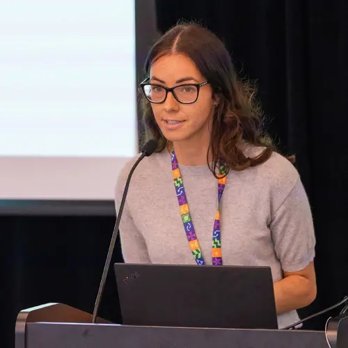
Portrait de Tal Arbel

Tal Arbel

Membre académique principal
Chaire en IA Canada-CIFAR
Professeure titulaire, McGill University, Département de génie électrique et informatique
Sujets de recherche
Apprentissage automatique médical
Apprentissage de représentations
Apprentissage profond
Causalité
Modèles génératifs
Modèles probabilistes
Vision par ordinateur

Biographie

Tal Arbel est professeure titulaire au Département de génie électrique et informatique de l’Université McGill, où elle dirige le groupe de vision probabiliste et le laboratoire d'imagerie médicale du Centre sur les machines intelligentes.

Elle est titulaire d'une chaire en IA Canada-CIFAR et membre associée de Mila – Institut québécois d’intelligence artificielle ainsi que du Centre de recherche sur le cancer Goodman. Les recherches de la professeure Arbel portent sur le développement de méthodes probabilistes d'apprentissage profond dans les domaines de la vision par ordinateur et de l’analyse d'imagerie médicale pour un large éventail d'applications dans le monde réel, avec un accent particulier sur les maladies neurologiques.

Elle a remporté le prix de la recherche Christophe Pierre 2019 de McGill Engineering. Elle fait régulièrement partie de l'équipe organisatrice de grandes conférences internationales sur la vision par ordinateur et l'analyse d'imagerie médicale (par exemple celles de la Medical Image Computing and Computer-Assisted Intervention Society/MICCAI et de Medical Imaging with Deep Learning/MIDL, l’International Conference on Computer Vision/ICCV ou encore la Conference on Computer Vision and Pattern Recognition/CVPR). Elle est rédactrice en chef et cofondatrice de la revue Machine Learning for Biomedical Imaging (MELBA).

Étudiants actuels

Postdoctorat - McGill
Doctorat - McGill
Maîtrise recherche - McGill
Doctorat - McGill
Maîtrise recherche - McGill
Baccalauréat - McGill

Publications

Combining intraoperative ultrasound brain shift correction and augmented reality visualizations: a pilot study of eight cases
Ian J. Gerard
Marta Kersten-Oertel
Simon Drouin
Jeffery A. Hall
Kevin Petrecca
Dante De Nigris
Daniel A. Di Giovanni
D. Louis Collins
Abstract. We present our work investigating the feasibility of combining intraoperative ultrasound for brain shift correction and augmented … (voir plus)reality (AR) visualization for intraoperative interpretation of patient-specific models in image-guided neurosurgery (IGNS) of brain tumors. We combine two imaging technologies for image-guided brain tumor neurosurgery. Throughout surgical interventions, AR was used to assess different surgical strategies using three-dimensional (3-D) patient-specific models of the patient’s cortex, vasculature, and lesion. Ultrasound imaging was acquired intraoperatively, and preoperative images and models were registered to the intraoperative data. The quality and reliability of the AR views were evaluated with both qualitative and quantitative metrics. A pilot study of eight patients demonstrates the feasible combination of these two technologies and their complementary features. In each case, the AR visualizations enabled the surgeon to accurately visualize the anatomy and pathology of interest for an extended period of the intervention. Inaccuracies associated with misregistration, brain shift, and AR were improved in all cases. These results demonstrate the potential of combining ultrasound-based registration with AR to become a useful tool for neurosurgeons to improve intraoperative patient-specific planning by improving the understanding of complex 3-D medical imaging data and prolonging the reliable use of IGNS.
Challenging Conventional Segmentation Evaluation Metrics Focal Pathology ( Lesion and Tumour ) Segmentation from Patient Images
How can we do better ? Pitfalls in biomedical challenge design and how to address them
Annika Reinke
Matthias Eisenmann
Sinan Onogur
Marko Stankovic
Patrick Scholz
Hrvoje Bogunovic
Andrew P. Bradley
Aaron
Carass
Carolin Feldmann
Alejandro F. Frangi
Peter M. Full
Bram Ginneken Van
Ginneken
Allan Hanbury
Katrin Honauer
Michal Kozubek
Adam Bennett
Landman … (voir 22 de plus)
Keno März
Oskar Maier
Klaus Maier-Hein
Bjoern Menze
Henning Müller
Peter F. Neher
Wiro Niessen
NASIR RAJPOOT
Catherine Gregory
Sharp
Korsuk Sirinukunwattana
Stefanie Speidel
Christian Stock
Danail
Stoyanov
Abdel Aziz Taha
F. V. D. Sommen
Ching-Wei Wang
Marc-André Weber
Guoyan Zheng
Pierre Jannin
Lena Maier-Hein
Since the first MICCAI grand challenge was organized in 2007 [1], the impact of biomedical image analysis challenges on both the research fi… (voir plus)eld as well as on individual careers has been steadily growing. For example, the acceptance of a journal article today often depends on the performance of a new algorithm being assessed against the state-ofthe-art work on publicly available challenge datasets. Furthermore, the results are also important for the individuals scientific careers as well as the potential that algorithms can be translated into clinical practice. Yet, while the publication of papers in scientific journals and prestigious conferences, such as MICCAI, undergoes strict quality control, the design and organization of challenges do not. To investigate the effect of common practice, we have formed an international initiative dedicated to analyzing and improving a variety of aspects related to biomedical challenge design, execution and reporting [2]. In the first part of our abstract presentation at LABELS workshop, we are going to present some of the major pitfalls related to biomedical image analysis challenges today. Specifically, we will look at the following research questions: RQ1: How robust are challenge rankings? What is the effect of – the specific test cases used? – the specific metric variant(s) applied? – the rank aggregation method chosen (e.g. aggregation of metric values with the mean vs median)? ? Shared first/senior authors.
Predicting Future Disease Activity and Treatment Responders for Multiple Sclerosis Patients Using a Bag-of-Lesions Brain Representation
Andrew Doyle
Douglas Arnold
Graphs in Biomedical Image Analysis, Computational Anatomy and Imaging Genetics
M. Cardoso
Enzo Ferrante
Xavier Pennec
Adrian Dalca
Sarah Parisot
S. Joshi
Nematollah Batmanghelich
Aristeidis Sotiras
Mads Lenstrup Nielsen
M. Sabuncu
Tom Fletcher
Li Shen
Stanley Durrleman
Stefan H. Sommer
Deep Learning in Medical Image Analysis and Multimodal Learning for Clinical Decision Support
M. Cardoso
G. Carneiro
T. Syeda-Mahmood
J. Tavares
Mehdi Moradi
Andrew P. Bradley
Hayit Greenspan
J. Papa
Anant. Madabhushi
Jacinto C Nascimento
Jaime S. Cardoso
Vasileios Belagiannis
Zhi Lu
Faculdade Engenharia
Computer Assisted and Robotic Endoscopy and Clinical Image-Based Procedures
M. Cardoso
Xiongbiao Luo
Stefan Wesarg
Tobias Reichl
M. Ballester
Jonathan Mcleod
Klaus Dr. Drechsler
T. Peters
Marius Erdt
Kensaku Mori
M. Linguraru
Andreas Uhl
Cristina Oyarzun Laura
R. Shekhar
Computer Assisted and Robotic Endoscopy and Clinical Image-Based Procedures
M. Jorge Cardoso
Xiongbiao Luo
Stefan Wesarg
Tobias Reichl
M. Ballester
Jonathan Mcleod
Klaus Dr. Drechsler
T. Peters
Marius Erdt
Kensaku Mori
M. Linguraru
Andreas Uhl
Cristina Oyarzun Laura
R. Shekhar
Deep Learning in Medical Image Analysis and Multimodal Learning for Clinical Decision Support
M. Jorge Cardoso
G. Carneiro
T. Syeda-Mahmood
J. Tavares
Mehdi Moradi
Andrew P. Bradley
Hayit Greenspan
J. Papa
Anant. Madabhushi
Jacinto C Nascimento
Jaime S. Cardoso
Vasileios Belagiannis
Zhi Lu
Faculdade Engenharia
Fetal, Infant and Ophthalmic Medical Image Analysis
M. Cardoso
Andrew Melbourne
Hrvoje Bogunovic
Pim Moeskops
Xinjian Chen
Ernst Schwartz
M. Garvin
E. Robinson
E. Trucco
Michael Ebner
Yanwu Xu
Antonios Makropoulos
Adrien Desjardin
Tom Kamiel Magda Vercauteren
Fetal, Infant and Ophthalmic Medical Image Analysis
M. Jorge Cardoso
Andrew Melbourne
Hrvoje Bogunovic
Pim Moeskops
Xinjian Chen
Ernst Schwartz
M. Garvin
E. Robinson
E. Trucco
Michael Ebner
Yanwu Xu
Antonios Makropoulos
Adrien Desjardin
Tom Kamiel Magda Vercauteren
Graphs in Biomedical Image Analysis, Computational Anatomy and Imaging Genetics
M. Jorge Cardoso
Enzo Ferrante
Xavier Pennec
Adrian Dalca
Sarah Parisot
S. Joshi
Nematollah Batmanghelich
Aristeidis Sotiras
Mads Lenstrup Nielsen
Mert R. Sabuncu
Tom Fletcher
Li Shen
Stanley Durrleman
Stefan H. Sommer