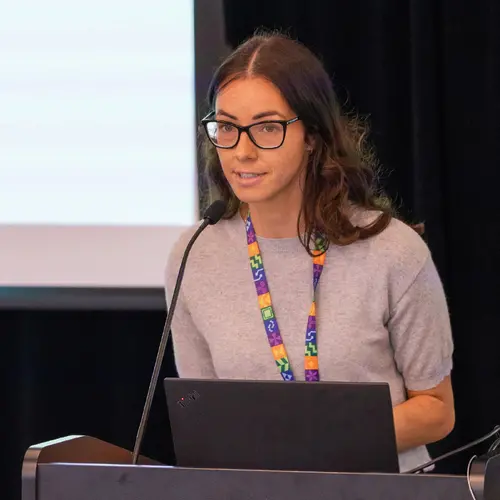
Portrait of Tal Arbel

Tal Arbel

Core Academic Member
Canada CIFAR AI Chair
Full Professor, McGill University, Department of Electrical and Computer Engineering
Research Topics
Causality
Computer Vision
Deep Learning
Generative Models
Medical Machine Learning
Probabilistic Models
Representation Learning

Biography

Tal Arbel is a professor in the Department of Electrical and Computer Engineering at McGill University, where she is the director of the Probabilistic Vision Group and Medical Imaging Lab in the Centre for Intelligent Machines.

She is also a Canada CIFAR AI Chair, an associate academic member of Mila – Quebec Artificial Intelligence Institute and an associate member of the Goodman Cancer Research Centre.

Arbel’s research focuses on the development of probabilistic deep learning methods in computer vision and medical image analysis for a wide range of real-world applications, with a focus on neurological diseases.

She is a recipient of the 2019 McGill Engineering Christophe Pierre Research Award and a Fellow of the Canadian Academy of Engineering. She regularly serves on the organizing team of major international conferences in computer vision and in medical image analysis (e.g. MICCAI, MIDL, ICCV, CVPR). She is currently the Editor-in-Chief and co-founder of the arXiv overlay journal: Machine Learning for Biomedical Imaging (MELBA).

Current Students

Collaborating researcher - Université de Montréal
Master's Research - McGill University
PhD - McGill University
Master's Research - McGill University
Undergraduate - McGill University
Master's Research - McGill University

Publications

Building a General SimCLR Self-Supervised Foundation Model Across Neurological Diseases to Advance 3D Brain MRI Diagnoses
3D structural Magnetic Resonance Imaging (MRI) brain scans are commonly acquired in clinical settings to monitor a wide range of neurologica… (see more)l conditions, including neurodegenerative disorders and stroke. While deep learning models have shown promising results analyzing 3D MRI across a number of brain imaging tasks, most are highly tailored for specific tasks with limited labeled data, and are not able to generalize across tasks and/or populations. The development of self-supervised learning (SSL) has enabled the creation of large medical foundation models that leverage diverse, unlabeled datasets ranging from healthy to diseased data, showing significant success in 2D medical imaging applications. However, even the very few foundation models for 3D brain MRI that have been developed remain limited in resolution, scope, or accessibility. In this work, we present a general, high-resolution SimCLR-based SSL foundation model for 3D brain structural MRI, pre-trained on 18,759 patients (44,958 scans) from 11 publicly available datasets spanning diverse neurological diseases. We compare our model to Masked Autoencoders (MAE), as well as two supervised baselines, on four diverse downstream prediction tasks in both in-distribution and out-of-distribution settings. Our fine-tuned SimCLR model outperforms all other models across all tasks. Notably, our model still achieves superior performance when fine-tuned using only 20% of labeled training samples for predicting Alzheimer's disease. We use publicly available code and data, and release our trained model at https://github.com/emilykaczmarek/3D-Neuro-SimCLR, contributing a broadly applicable and accessible foundation model for clinical brain MRI analysis.
SSL-AD: Spatiotemporal Self-Supervised Learning for Generalizability and Adaptability Across Alzheimer's Prediction Tasks and Datasets
Alzheimer's disease is a progressive, neurodegenerative disorder that causes memory loss and cognitive decline. While there has been extensi… (see more)ve research in applying deep learning models to Alzheimer's prediction tasks, these models remain limited by lack of available labeled data, poor generalization across datasets, and inflexibility to varying numbers of input scans and time intervals between scans. In this study, we adapt three state-of-the-art temporal self-supervised learning (SSL) approaches for 3D brain MRI analysis, and add novel extensions designed to handle variable-length inputs and learn robust spatial features. We aggregate four publicly available datasets comprising 3,161 patients for pre-training, and show the performance of our model across multiple Alzheimer's prediction tasks including diagnosis classification, conversion detection, and future conversion prediction. Importantly, our SSL model implemented with temporal order prediction and contrastive learning outperforms supervised learning on six out of seven downstream tasks. It demonstrates adaptability and generalizability across tasks and number of input images with varying time intervals, highlighting its capacity for robust performance across clinical applications. We release our code and model publicly at https://github.com/emilykaczmarek/SSL-AD.
Imagining Alternatives: Towards High-Resolution 3D Counterfactual Medical Image Generation via Language Guidance
Vision-language models have demonstrated impressive capabilities in generating 2D images under various conditions; however the impressive pe… (see more)rformance of these models in 2D is largely enabled by extensive, readily available pretrained foundation models. Critically, comparable pretrained foundation models do not exist for 3D, significantly limiting progress in this domain. As a result, the potential of vision-language models to produce high-resolution 3D counterfactual medical images conditioned solely on natural language descriptions remains completely unexplored. Addressing this gap would enable powerful clinical and research applications, such as personalized counterfactual explanations, simulation of disease progression scenarios, and enhanced medical training by visualizing hypothetical medical conditions in realistic detail. Our work takes a meaningful step toward addressing this challenge by introducing a framework capable of generating high-resolution 3D counterfactual medical images of synthesized patients guided by free-form language prompts. We adapt state-of-the-art 3D diffusion models with enhancements from Simple Diffusion and incorporate augmented conditioning to improve text alignment and image quality. To our knowledge, this represents the first demonstration of a language-guided native-3D diffusion model applied specifically to neurological imaging data, where faithful three-dimensional modeling is essential to represent the brain's three-dimensional structure. Through results on two distinct neurological MRI datasets, our framework successfully simulates varying counterfactual lesion loads in Multiple Sclerosis (MS), and cognitive states in Alzheimer's disease, generating high-quality images while preserving subject fidelity in synthetically generated medical images. Our results lay the groundwork for prompt-driven disease progression analysis within 3D medical imaging.
Pixels Under Pressure: Exploring Fine-Tuning Paradigms for Foundation Models in High-Resolution Medical Imaging
Zahra Tehrani Nasab
Advancements in diffusion-based foundation models have improved text-to-image generation, yet most efforts have been limited to low-resoluti… (see more)on settings. As high-resolution image synthesis becomes increasingly essential for various applications, particularly in medical imaging domains, fine-tuning emerges as a crucial mechanism for adapting these powerful pre-trained models to task-specific requirements and data distributions. In this work, we present a systematic study, examining the impact of various fine-tuning techniques on image generation quality when scaling to high resolution 512x512 pixels. We benchmark a diverse set of fine-tuning methods, including full fine-tuning strategies and parameter-efficient fine-tuning (PEFT). We dissect how different fine-tuning methods influence key quality metrics, including Fr\'echet Inception Distance (FID), Vendi score, and prompt-image alignment. We also evaluate the utility of generated images in a downstream classification task under data-scarce conditions, demonstrating that specific fine-tuning strategies improve both generation fidelity and downstream performance when synthetic images are used for classifier training and evaluation on real images. Our code is accessible through the project website - https://tehraninasab.github.io/PixelUPressure/.
Spatio-Temporal Conditional Diffusion Models for Forecasting Future Multiple Sclerosis Lesion Masks Conditioned on Treatments
Gian Mario Favero
Ge Ya Luo
Douglas Arnold
Image-based personalized medicine has the potential to transform healthcare, particularly for diseases that exhibit heterogeneous progressio… (see more)n such as Multiple Sclerosis (MS). In this work, we introduce the first treatment-aware spatio-temporal diffusion model that is able to generate future masks demonstrating lesion evolution in MS. Our voxel-space approach incorporates multi-modal patient data, including MRI and treatment information, to forecast new and enlarging T2 (NET2) lesion masks at a future time point. Extensive experiments on a multi-centre dataset of 2131 patient 3D MRIs from randomized clinical trials for relapsing-remitting MS demonstrate that our generative model is able to accurately predict NET2 lesion masks for patients across six different treatments. Moreover, we demonstrate our model has the potential for real-world clinical applications through downstream tasks such as future lesion count and location estimation, binary lesion activity classification, and generating counterfactual future NET2 masks for several treatments with different efficacies. This work highlights the potential of causal, image-based generative models as powerful tools for advancing data-driven prognostics in MS.
Spatio-Temporal Conditional Diffusion Models for Forecasting Future Multiple Sclerosis Lesion Masks Conditioned on Treatments
Gian Mario Favero
Ge Ya Luo
Douglas Arnold
Pixels Under Pressure: Exploring Fine-Tuning Paradigms for Foundation Models in High-Resolution Medical Imaging
Zahra Tehrani Nasab
Advancements in diffusion-based foundation models have improved text-to-image generation, yet most efforts have been limited to low-resoluti… (see more)on settings. As high-resolution image synthesis becomes increasingly essential for various applications, particularly in medical imaging domains, fine-tuning emerges as a crucial mechanism for adapting these powerful pre-trained models to task-specific requirements and data distributions. In this work, we present a systematic study, examining the impact of various fine-tuning techniques on image generation quality when scaling to high resolution 512x512 pixels. We benchmark a diverse set of fine-tuning methods, including full fine-tuning strategies and parameter-efficient fine-tuning (PEFT). We dissect how different fine-tuning methods influence key quality metrics, including Fréchet Inception Distance (FID), Vendi score, and prompt-image alignment. We also evaluate the utility of generated images in a downstream classification task under data-scarce conditions, demonstrating that specific fine-tuning strategies improve both generation fidelity and downstream performance when synthetic images are used for classifier training and evaluation on real images. Our code is accessible through the project website - https://tehraninasab.github.io/PixelUPressure/.
Spatio-Temporal Conditional Diffusion Models for Forecasting Future Multiple Sclerosis Lesion Masks Conditioned on Treatments
Gian Mario Favero
Ge Ya Luo
Douglas Arnold
Image-based personalized medicine has the potential to transform healthcare, particularly for diseases that exhibit heterogeneous progressio… (see more)n such as Multiple Sclerosis (MS). In this work, we introduce the first treatment-aware spatio-temporal diffusion model that is able to generate future masks demonstrating lesion evolution in MS. Our voxel-space approach incorporates multi-modal patient data, including MRI and treatment information, to forecast new and enlarging T2 (NET2) lesion masks at a future time point. Extensive experiments on a multi-centre dataset of 2131 patient 3D MRIs from randomized clinical trials for relapsing-remitting MS demonstrate that our generative model is able to accurately predict NET2 lesion masks for patients across six different treatments. Moreover, we demonstrate our model has the potential for real-world clinical applications through downstream tasks such as future lesion count and location estimation, binary lesion activity classification, and generating counterfactual future NET2 masks for several treatments with different efficacies. This work highlights the potential of causal, image-based generative models as powerful tools for advancing data-driven prognostics in MS.
AURA: A Multi-Modal Medical Agent for Understanding, Reasoning&Annotation
Recent advancements in Large Language Models (LLMs) have catalyzed a paradigm shift from static prediction systems to agentic AI agents capa… (see more)ble of reasoning, interacting with tools, and adapting to complex tasks. While LLM-based agentic systems have shown promise across many domains, their application to medical imaging remains in its infancy. In this work, we introduce AURA, the first visual linguistic explainability agent designed specifically for comprehensive analysis, explanation, and evaluation of medical images. By enabling dynamic interactions, contextual explanations, and hypothesis testing, AURA represents a significant advancement toward more transparent, adaptable, and clinically aligned AI systems. We highlight the promise of agentic AI in transforming medical image analysis from static predictions to interactive decision support. Leveraging Qwen-32B, an LLM-based architecture, AURA integrates a modular toolbox comprising: (i) a segmentation suite with phase grounding, pathology segmentation, and anatomy segmentation to localize clinically meaningful regions; (ii) a counterfactual image-generation module that supports reasoning through image-level explanations; and (iii) a set of evaluation tools including pixel-wise difference-map analysis, classification, and advanced state-of-the-art components to assess diagnostic relevance and visual interpretability.