Événement
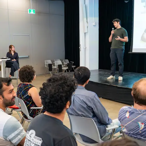
Science éclair
Rejoignez-nous le 19 novembre pour la troisième édition du concours de vulgarisation scientifique de Mila, où les étudiant·e·s présenteront leurs recherches complexes en trois minutes devant un jury.
Rapport d'impact 2024-2025
À l’avant-garde d’une nouvelle ère
Le dernier rapport d'impact de Mila met en lumière les réalisations exceptionnelles des membres de notre communauté au cours de la dernière année.
Futurs étudiants / Postdocs
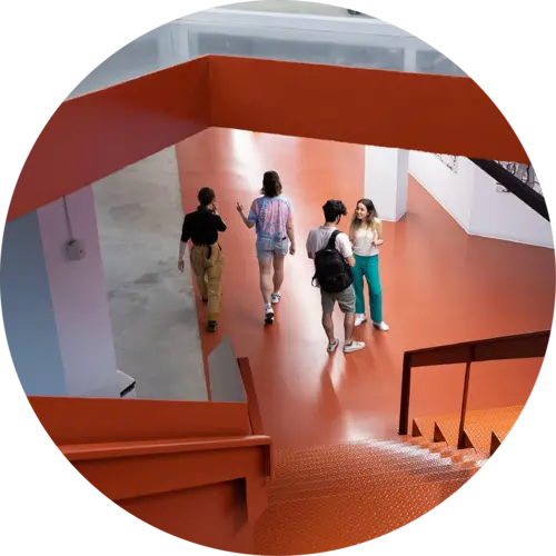
Demandes de supervision
Le processus de demande de supervision de Mila pour les admissions à l’automne 2026 est maintenant ouvert jusqu'au 1er décembre.