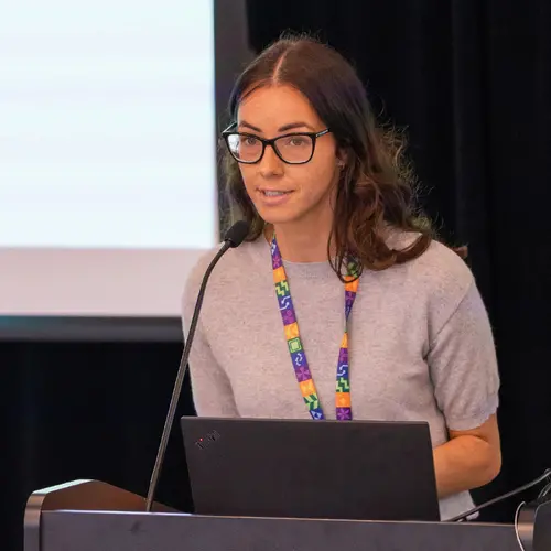
Portrait of Martin Vallières

Martin Vallières

Associate Academic Member
Canada CIFAR AI Chair
Assistant Professor, Université Sherbrooke, Department of Computer Science
Research Topics
Medical Machine Learning

Biography

Martin Vallières is an assistant professor in the Department of Computer Science at Université de Sherbrooke and a Canada CIFAR AI Chair since April 2020.

He received a PhD in medical physics from McGill University in 2017, and completed postdoctoral training in France and the U.S. in 2018 and 2019.

Vallières is an expert in the field of radiomics and machine learning in oncology. Over the course of his career, he has developed multiple prediction models for different types of cancers. His main research interest is now focused on the graph-based modelling of heterogeneous medical data for improved precision medicine.

Current Students

PhD - Université de Sherbrooke
Principal supervisor :
PhD - Université de Sherbrooke
Principal supervisor :

Publications

Towards fair decentralized benchmarking of healthcare AI algorithms with the Federated Tumor Segmentation (FeTS) challenge
Maximilian Zenk
Ujjwal Baid
Sarthak Pati
Akis Linardos
Brandon Edwards
Micah Sheller
Patrick Foley
Alejandro Aristizabal
David Zimmerer
Alexey Gruzdev
Jason Martin
Russell T. Shinohara
Annika Reinke
Fabian Isensee
Santhosh Parampottupadam
Kaushal Parekh
Ralf Floca
Hasan Kassem
Bhakti Baheti
Siddhesh Thakur … (see 332 more)
Verena Chung
Kaisar Kushibar
Karim Lekadir
Meirui Jiang
Youtan Yin
Hongzheng Yang
Quande Liu
Cheng Chen
Qi Dou
Pheng-Ann Heng
Xiaofan Zhang
Shaoting Zhang
Muhammad Irfan Khan
Mohammad Ayyaz Azeem
Mojtaba Jafaritadi
Esa Alhoniemi
Elina Kontio
Suleiman A. Khan
Leon Mächler
Ivan Ezhov
Florian Kofler
Suprosanna Shit
Johannes C. Paetzold
Timo Loehr
Benedikt Wiestler
Himashi Peiris
Kamlesh Pawar
Shenjun Zhong
Zhaolin Chen
Munawar Hayat
Gary Egan
Mehrtash Harandi
Ece Isik Polat
Gorkem Polat
Altan Kocyigit
Alptekin Temizel
Anup Tuladhar
Lakshay Tyagi
Raissa Souza
Nils D. Forkert
Pauline Mouches
Matthias Wilms
Vishruth Shambhat
Akansh Maurya
Shubham Subhas Danannavar
Rohit Kalla
Vikas Kumar Anand
Ganapathy Krishnamurthi
Sahil Nalawade
Chandan Ganesh
Ben Wagner
Divya Reddy
Yudhajit Das
Fang F. Yu
Baowei Fei
B. Fei
Ananth J. Madhuranthakam
Joseph Maldjian
Gaurav Singh
Jianxun Ren
Wei Zhang
Ning An
Qingyu Hu
Youjia Zhang
Ying Zhou
Vasilis Siomos
Giacomo Tarroni
Jonathan Passerrat-Palmbach
Ambrish Rawat
Giulio Zizzo
Swanand Ravindra Kadhe
Jonathan P. Epperlein
Stefano Braghin
Yuan Wang
Renuga Kanagavelu
Qingsong Wei
Yechao Yang
Yong Liu
Krzysztof Kotowski
Szymon Adamski
Bartosz Machura
Wojciech Malara
Lukasz Zarudzki
Jakub Nalepa
Yaying Shi
Hongjian Gao
Salman Avestimehr
Yonghong Yan
Agus S. Akbar
Ekaterina Kondrateva
Hua Yang
Zhaopei Li
Hung-Yu Wu
Johannes Roth
Camillo Saueressig
Alexandre Milesi
Quoc D. Nguyen
Nathan J. Gruenhagen
Tsung-Ming Huang
Jun Ma
Har Shwinder H. Singh
Nai-Yu Pan
Dingwen Zhang
Ramy A. Zeineldin
Michal Futrega
Yading Yuan
Gian Marco Conte
GM Conte
Xue Feng
Quan D. Pham
Yong Xia
Zhifan Jiang
Huan Minh Luu
Mariia Dobko
Alexandre Carré
Bair Tuchinov
Hassan Mohy-ud-Din
Saruar Alam
Anup Singh
Nameeta Shah
Weichung Wang
Chiharu Sako
Michel Bilello
Satyam Ghodasara
Suyash Mohan
Christos Davatzikos
Evan Calabrese
Jeffrey Rudie
Javier Villanueva-Meyer
S. Cha
Soonmee Cha
Christopher Hess
John Mongan
Madhura Ingalhalikar
Manali Jadhav
Umang Pandey
Jitender Saini
Raymond Y. Huang
Ken Chang
Minh-Son To
Sargam Bhardwaj
Chee Chong
Marc Agzarian
Michal Kozubek
Filip Lux
Jan Michálek
Petr Matula
Miloš Ker^kovský
Tereza Kopr^ivová
Marek Dostál
Václav Vybíhal
Marco C. Pinho
James Holcomb
Marie Metz
Rajan Jain
Matthew D. Lee
Yvonne W. Lui
Pallavi Tiwari
Ruchika Verma
Rohan Bareja
Ipsa Yadav
Jonathan Chen
Neeraj Kumar
Yuriy Gusev
Krithika Bhuvaneshwar
Anousheh Sayah
Camelia Bencheqroun
Anas Belouali
Subha Madhavan
Rivka R. Colen
Aikaterini Kotrotsou
Philipp Vollmuth
Gianluca Brugnara
Chandrakanth J. Preetha
Felix Sahm
Martin Bendszus
Wolfgang Wick
Abhishek Mahajan
Carmen Balaña
Jaume Capellades
Josep Puig
Yoon Seong Choi
Seung-Koo Lee
Jong Hee Chang
Sung Soo Ahn
Hassan F. Shaykh
Alejandro Herrera-Trujillo
Maria Trujillo
William Escobar
Ana Abello
Jose Bernal
Jhon Gómez
Pamela LaMontagne
Daniel S. Marcus
Mikhail Milchenko
Arash Nazeri
BENNETT A. LANDMAN
Karthik Ramadass
Kaiwen Xu
Silky Chotai
Lola B. Chambless
Akshitkumar Mistry
Reid C. Thompson
Ashok Srinivasan
Jayapalli R. Bapuraj
J. Rajiv Bapuraj
Arvind Rao
Nicholas Wang
Ota Yoshiaki
Toshio Moritani
Sevcan Turk
Joonsang Lee
Snehal Prabhudesai
John Garrett
Matthew Larson
Robert Jeraj
Hongwei Li
H. Li
Tobias Weiss
Michael Weller
Andrea Bink
Bertrand Pouymayou
Sonam Sharma
Tzu-Chi Tseng
Saba Adabi
Alexandre Xavier Falcão
Samuel B. Martins
Bernardo C. A. Teixeira
Flávia Sprenger
David Menotti
Diego R. Lucio
Simone P. Niclou
Olivier Keunen
Ann-Christin Hau
Enrique Pelaez
Heydy Franco-Maldonado
Francis Loayza
Sebastian Quevedo
Richard McKinley
Johannes Slotboom
Piotr Radojewski
Raphael Meier
Roland Wiest
Johannes Trenkler
Josef Pichler
Georg Necker
Andreas Haunschmidt
Stephan Meckel
Pamela Guevara
Esteban Torche
Cristobal Mendoza
Franco Vera
Elvis Ríos
Eduardo López
Sergio A. Velastin
Joseph Choi
Stephen Baek
Yusung Kim
Heba Ismael
Bryan Allen
John M. Buatti
Peter Zampakis
Vasileios Panagiotopoulos
Panagiotis Tsiganos
Sotiris Alexiou
Ilias Haliassos
Evangelia I. Zacharaki
Konstantinos Moustakas
Christina Kalogeropoulou
Dimitrios M. Kardamakis
Bing Luo
Laila M. Poisson
Ning Wen
Mahdi A. L. Loutfi
David Fortin
Martin Lepage
Fanny Morón
Jacob Mandel
Gaurav Shukla
Spencer Liem
Gregory S. Alexandre
Joseph Lombardo
Joshua D. Palmer
Adam E. Flanders
Adam P. Dicker
Godwin Ogbole
Dotun Oyekunle
Olubunmi Odafe-Oyibotha
Babatunde Osobu
Mustapha Shu’aibu Hikima
Mayowa Soneye
Farouk Dako
Adeleye Dorcas
Derrick Murcia
Eric Fu
Rourke Haas
John A. Thompson
David Ryan Ormond
Stuart Currie
Kavi Fatania
Russell Frood
Amber L. Simpson
Jacob J. Peoples
Ricky Hu
Danielle Cutler
Fabio Y. Moraes
Anh Tran
Mohammad Hamghalam
Michael A. Boss
James Gimpel
Deepak Kattil Veettil
Kendall Schmidt
Lisa Cimino
Cynthia Price
Brian Bialecki
Sailaja Marella
Charles Apgar
Andras Jakab
Marc-André Weber
Errol Colak
Jens Kleesiek
John Freymann
Justin Kirby
Lena Maier-Hein
Jake Albrecht
Peter Mattson
Alexandros Karargyris
Prashant Shah
Bjoern Menze
Klaus Maier-Hein
Spyridon Bakas
Computational competitions are the standard for benchmarking medical image analysis algorithms, but they typically use small curated test da… (see more)tasets acquired at a few centers, leaving a gap to the reality of diverse multicentric patient data. To this end, the Federated Tumor Segmentation (FeTS) Challenge represents the paradigm for real-world algorithmic performance evaluation. The FeTS challenge is a competition to benchmark (i) federated learning aggregation algorithms and (ii) state-of-the-art segmentation algorithms, across multiple international sites. Weight aggregation and client selection techniques were compared using a multicentric brain tumor dataset in realistic federated learning simulations, yielding benefits for adaptive weight aggregation, and efficiency gains through client sampling. Quantitative performance evaluation of state-of-the-art segmentation algorithms on data distributed internationally across 32 institutions yielded good generalization on average, albeit the worst-case performance revealed data-specific modes of failure. Similar multi-site setups can help validate the real-world utility of healthcare AI algorithms in the future.
Derivation and validation of indices incorporating vasopressor dose and blood pressure values over time
Alain Gervais
François Lamontagne
Jean-Baptiste Michaud
KJ Neill
Adhikari
Jean-Michel Pagé
Marie-Hélène Masse
Michael O. Harhay
Michael Chassé
Félix Lamontagne
Katia Laforge
Alexandra Fortin
Marc-André Leclair
Simon Lévesque
Marie-Pier Domingue
Neda Momenzadeh
Ruxandra Pinto
Maxime Morin-Lavoie
Francis Carter … (see 2 more)
Félix Camirand Lemyre
MD MSc. François Lamontagne
Rationale The blood pressure value below which the benefits of vasopressors clearly outweigh their disadvantages is uncertain. Objectives Th… (see more)e main objective of this analysis was to investigate the statistical properties and potential utility of indices estimating the vasopressor dose-rates as a function of blood pressure values over time. Methods In this single-center observational study, we collected blood pressure values from intensive care unit (ICU) monitors and norepinephrine dose-rates from infusion pumps corresponding to a derivation and a validation cohort. Patients included in each cohort were 18 years or older and received norepinephrine in the ICU. We defined and derived indices corresponding to vasopressor therapy above (>65 mmHg) and below (60 mmHg) targets. We report the distribution of both indices over time from both cohorts as well as their associations with hospital mortal
Derivation and validation of indices incorporating vasopressor dose and blood pressure values over time
Alain Gervais
François Lamontagne
Jean-Baptiste Michaud
KJ Neill
Adhikari
Jean-Michel Pagé
Marie-Hélène Masse
Michael O Harhay
Michael Chassé
Félix Lamontagne
Katia Laforge
Alexandra Fortin
Marc-André Leclair
Simon Lévesque
Marie-Pier Domingue
Neda Momenzadeh
Ruxandra Pinto
Maxime Morin-Lavoie
Francis Carter … (see 2 more)
Félix Camirand Lemyre
MD MSc. François Lamontagne
Rationale The blood pressure value below which the benefits of vasopressors clearly outweigh their disadvantages is uncertain. Objectives Th… (see more)e main objective of this analysis was to investigate the statistical properties and potential utility of indices estimating the vasopressor dose-rates as a function of blood pressure values over time. Methods In this single-center observational study, we collected blood pressure values from intensive care unit (ICU) monitors and norepinephrine dose-rates from infusion pumps corresponding to a derivation and a validation cohort. Patients included in each cohort were 18 years or older and received norepinephrine in the ICU. We defined and derived indices corresponding to vasopressor therapy above (>65 mmHg) and below (60 mmHg) targets. We report the distribution of both indices over time from both cohorts as well as their associations with hospital mortal
Development of Error Passing Network for Optimizing the Prediction of VO$_2$ peak in Childhood Acute Leukemia Survivors
Nicolas Raymond
Hakima Laribi
Maxime Caru
Mehdi Mitiche
Valerie Marcil
Maja Krajinovic
Daniel Curnier
Daniel Sinnett
Approximately two-thirds of survivors of childhood acute lymphoblastic leukemia (ALL) cancer develop late adverse effects post-treatment. Pr… (see more)ior studies explored prediction models for personalized follow-up, but none integrated the usage of neural networks to date. In this work, we propose the Error Passing Network (EPN), a graph-based method that leverages relationships between samples to propagate residuals and adjust predictions of any machine learning model. We tested our approach to estimate patients’ \vo peak, a reliable indicator of their cardiac health. We used the EPN in conjunction with several baseline models and observed up to 12.16% improvement in the mean average percentage error compared to the last established equation predicting \vo peak in childhood ALL survivors. Along with this performance improvement, our final model is more efficient considering that it relies only on clinical variables that can be self-reported by patients, therefore removing the previous need of executing a resource-consuming physical test.
Unraveling Radiomics Complexity: Strategies for Optimal Simplicity in Predictive Modeling
Mahdi A. L. Loutfi
Teodora Boblea Podasca
Alex Zwanenburg
Taman Upadhaya
Jorge Barrios
David R Raleigh
William C. Chen
Dante P. I. Capaldi
Hong Zheng
Olivier Gevaert
Jinglan Wu
Alvin C. Silva
Paul J. Zhang
Harrison X. Bai
Jan Seuntjens
Steffen Löck
Patrick O. Richard
Olivier Morin
Caroline Reinhold
Martin Lepage … (see 1 more)
Background: The high dimensionality of radiomic feature sets, the variability in radiomic feature types and potentially high computational r… (see more)equirements all underscore the need for an effective method to identify the smallest set of predictive features for a given clinical problem. Purpose: Develop a methodology and tools to identify and explain the smallest set of predictive radiomic features. Materials and Methods: 89,714 radiomic features were extracted from five cancer datasets: low-grade glioma, meningioma, non-small cell lung cancer (NSCLC), and two renal cell carcinoma cohorts (n=2104). Features were categorized by computational complexity into morphological, intensity, texture, linear filters, and nonlinear filters. Models were trained and evaluated on each complexity level using the area under the curve (AUC). The most informative features were identified, and their importance was explained. The optimal complexity level and associated most informative features were identified using systematic statistical significance analyses and a false discovery avoidance procedure, respectively. Their predictive importance was explained using a novel tree-based method. Results: MEDimage, a new open-source tool, was developed to facilitate radiomic studies. Morphological features were optimal for MRI-based meningioma (AUC: 0.65) and low-grade glioma (AUC: 0.68). Intensity features were optimal for CECT-based renal cell carcinoma (AUC: 0.82) and CT-based NSCLC (AUC: 0.76). Texture features were optimal for MRI-based renal cell carcinoma (AUC: 0.72). Tuning the Hounsfield unit range improved results for CECT-based renal cell carcinoma (AUC: 0.86). Conclusion: Our proposed methodology and software can estimate the optimal radiomics complexity level for specific medical outcomes, potentially simplifying the use of radiomics in predictive modeling across various contexts.
The Image Biomarker Standardization Initiative: Standardized Convolutional Filters for Reproducible Radiomics and Enhanced Clinical Insights.
Philip Whybra
Alex Zwanenburg
Vincent Andrearczyk
Roger Schaer
Aditya P. Apte
Alexandre Ayotte
Bhakti Baheti
Spyridon Bakas
Andrea Bettinelli
Ronald Boellaard
Luca Boldrini
Irene Buvat
Gary J. R. Cook
Florian Dietsche
Nicola Dinapoli
Hubert S. Gabryś
Vicky Goh
Matthias Guckenberger
Mathieu Hatt
Mahdi Hosseinzadeh … (see 26 more)
Aditi Iyer
Jacopo Lenkowicz
Mahdi A. L. Loutfi
Steffen Löck
Francesca Marturano
Olivier Morin
Christophe Nioche
Fanny Orlhac
Sarthak Pati
Arman Rahmim
Seyed Masoud Rezaeijo
Christopher G. Rookyard
Mohammad R. Salmanpour
Andreas Schindele
Isaac Shiri
Emiliano Spezi
Stephanie Tanadini-Lang
Florent Tixier
Taman Upadhaya
Vincenzo Valentini
Joost J. M. van Griethuysen
Fereshteh Yousefirizi
Habib Zaidi
Henning Müller
Adrien Depeursinge
Filters are commonly used to enhance specific structures and patterns in images, such as vessels or peritumoral regions, to enable clinical … (see more)insights beyond the visible image using radiomics. However, their lack of standardization restricts reproducibility and clinical translation of radiomics decision support tools. In this special report, teams of researchers who developed radiomics software participated in a three-phase study (September 2020 to December 2022) to establish a standardized set of filters. The first two phases focused on finding reference filtered images and reference feature values for commonly used convolutional filters: mean, Laplacian of Gaussian, Laws and Gabor kernels, separable and nonseparable wavelets (including decomposed forms), and Riesz transformations. In the first phase, 15 teams used digital phantoms to establish 33 reference filtered images of 36 filter configurations. In phase 2, 11 teams used a chest CT image to derive reference values for 323 of 396 features computed from filtered images using 22 filter and image processing configurations. Reference filtered images and feature values for Riesz transformations were not established. Reproducibility of standardized convolutional filters was validated on a public data set of multimodal imaging (CT, fluorodeoxyglucose PET, and T1-weighted MRI) in 51 patients with soft-tissue sarcoma. At validation, reproducibility of 486 features computed from filtered images using nine configurations × three imaging modalities was assessed using the lower bounds of 95% CIs of intraclass correlation coefficients. Out of 486 features, 458 were found to be reproducible across nine teams with lower bounds of 95% CIs of intraclass correlation coefficients greater than 0.75. In conclusion, eight filter types were standardized with reference filtered images and reference feature values for verifying and calibrating radiomics software packages. A web-based tool is available for compliance checking.
The Image Biomarker Standardization Initiative: Standardized Convolutional Filters for Reproducible Radiomics and Enhanced Clinical Insights
Philip Whybra
Alex Zwanenburg
Vincent Andrearczyk
Roger Schaer
Aditya P. Apte
Alexandre Ayotte
Bhakti Baheti
Spyridon Bakas
Andrea Bettinelli
Ronald Boellaard
Luca Boldrini
Irene Buvat
Gary J. R. Cook
Florian Dietsche
Nicola Dinapoli
Hubert S. Gabryś
Vicky Goh
Matthias Guckenberger
Mathieu Hatt
Mahdi Hosseinzadeh … (see 26 more)
Aditi Iyer
Jacopo Lenkowicz
Mahdi A. L. Loutfi
Steffen Löck
Francesca Marturano
Olivier Morin
Christophe Nioche
Fanny Orlhac
Sarthak Pati
Arman Rahmim
Seyed Masoud Rezaeijo
Christopher G. Rookyard
Mohammad R. Salmanpour
Andreas Schindele
Isaac Shiri
Emiliano Spezi
Stephanie Tanadini-Lang
Florent Tixier
Taman Upadhaya
Vincenzo Valentini
Joost J. M. van Griethuysen
Fereshteh Yousefirizi
Habib Zaidi
Henning Müller
Adrien Depeursinge
The Image Biomarker Standardization Initiative: Standardized Convolutional Filters for Reproducible Radiomics and Enhanced Clinical Insights.
Philip Whybra
Alex Zwanenburg
Vincent Andrearczyk
Roger Schaer
Aditya P. Apte
Alexandre Ayotte
Bhakti Baheti
Spyridon Bakas
Andrea Bettinelli
Ronald Boellaard
Luca Boldrini
Irene Buvat
Gary J. R. Cook
Florian Dietsche
Nicola Dinapoli
Hubert S. Gabryś
Vicky Goh
Matthias Guckenberger
Mathieu Hatt
Mahdi Hosseinzadeh … (see 26 more)
Aditi Iyer
Jacopo Lenkowicz
Mahdi A. L. Loutfi
Steffen Löck
Francesca Marturano
Olivier Morin
Christophe Nioche
Fanny Orlhac
Sarthak Pati
Arman Rahmim
Seyed Masoud Rezaeijo
Christopher G. Rookyard
Mohammad R. Salmanpour
Andreas Schindele
Isaac Shiri
Emiliano Spezi
Stephanie Tanadini-Lang
Florent Tixier
Taman Upadhaya
Vincenzo Valentini
Joost J. M. van Griethuysen
Fereshteh Yousefirizi
Habib Zaidi
Henning Müller
Adrien Depeursinge
Filters are commonly used to enhance specific structures and patterns in images, such as vessels or peritumoral regions, to enable clinical … (see more)insights beyond the visible image using radiomics. However, their lack of standardization restricts reproducibility and clinical translation of radiomics decision support tools. In this special report, teams of researchers who developed radiomics software participated in a three-phase study (September 2020 to December 2022) to establish a standardized set of filters. The first two phases focused on finding reference filtered images and reference feature values for commonly used convolutional filters: mean, Laplacian of Gaussian, Laws and Gabor kernels, separable and nonseparable wavelets (including decomposed forms), and Riesz transformations. In the first phase, 15 teams used digital phantoms to establish 33 reference filtered images of 36 filter configurations. In phase 2, 11 teams used a chest CT image to derive reference values for 323 of 396 features computed from filtered images using 22 filter and image processing configurations. Reference filtered images and feature values for Riesz transformations were not established. Reproducibility of standardized convolutional filters was validated on a public data set of multimodal imaging (CT, fluorodeoxyglucose PET, and T1-weighted MRI) in 51 patients with soft-tissue sarcoma. At validation, reproducibility of 486 features computed from filtered images using nine configurations × three imaging modalities was assessed using the lower bounds of 95% CIs of intraclass correlation coefficients. Out of 486 features, 458 were found to be reproducible across nine teams with lower bounds of 95% CIs of intraclass correlation coefficients greater than 0.75. In conclusion, eight filter types were standardized with reference filtered images and reference feature values for verifying and calibrating radiomics software packages. A web-based tool is available for compliance checking.