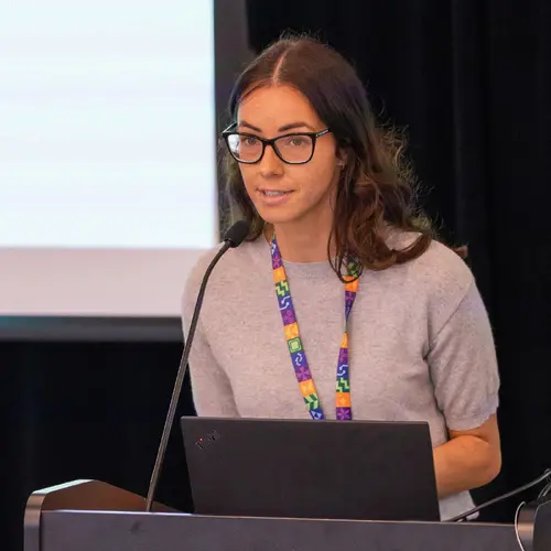
Portrait de Martin Vallières

Martin Vallières

Membre académique associé
Chaire en IA Canada-CIFAR
Professeur adjoint, Université Sherbrooke, Département d'informatique
Sujets de recherche
Apprentissage automatique médical

Biographie

Martin Vallières est professeur adjoint au Département d'informatique de l'Université de Sherbrooke et titulaire d'une chaire en IA Canada-CIFAR depuis avril 2020. Il a obtenu un doctorat en physique médicale de l'Université McGill en 2017, et a suivi une formation postdoctorale en France et aux États-Unis en 2018 et 2019. Il est expert dans le domaine de la radiomique et de l'apprentissage automatique en oncologie. Au cours de sa carrière, il a développé de multiples modèles de prédiction pour différents types de cancer. Son principal intérêt de recherche consiste désormais en la modélisation à base de graphes de données médicales hétérogènes pour améliorer la médecine de précision.

Étudiants actuels

Doctorat - Université de Sherbrooke
Superviseur⋅e principal⋅e :
Doctorat - Université de Sherbrooke
Superviseur⋅e principal⋅e :

Publications

Development of Error Passing Network for Optimizing the Prediction of VO$_2$ peak in Childhood Acute Leukemia Survivors
Nicolas Raymond
Hakima Laribi
Maxime Caru
Mehdi Mitiche
Valerie Marcil
Maja Krajinovic
Daniel Curnier
Daniel Sinnett
Approximately two-thirds of survivors of childhood acute lymphoblastic leukemia (ALL) cancer develop late adverse effects post-treatment. Pr… (voir plus)ior studies explored prediction models for personalized follow-up, but none integrated the usage of neural networks to date. In this work, we propose the Error Passing Network (EPN), a graph-based method that leverages relationships between samples to propagate residuals and adjust predictions of any machine learning model. We tested our approach to estimate patients’ \vo peak, a reliable indicator of their cardiac health. We used the EPN in conjunction with several baseline models and observed up to 12.16% improvement in the mean average percentage error compared to the last established equation predicting \vo peak in childhood ALL survivors. Along with this performance improvement, our final model is more efficient considering that it relies only on clinical variables that can be self-reported by patients, therefore removing the previous need of executing a resource-consuming physical test.
Unraveling Radiomics Complexity: Strategies for Optimal Simplicity in Predictive Modeling
Mahdi A. L. Loutfi
Teodora Boblea Podasca
Alex Zwanenburg
Taman Upadhaya
Jorge Barrios
David R Raleigh
William C. Chen
Dante P. I. Capaldi
Hong Zheng
Olivier Gevaert
Jing Wu
Alvin C. Silva
Paul J. Zhang
Harrison X. Bai
Jan Seuntjens
Steffen Löck
Patrick O. Richard
Olivier Morin
Caroline Reinhold
Martin Lepage … (voir 1 de plus)
Background: The high dimensionality of radiomic feature sets, the variability in radiomic feature types and potentially high computational r… (voir plus)equirements all underscore the need for an effective method to identify the smallest set of predictive features for a given clinical problem. Purpose: Develop a methodology and tools to identify and explain the smallest set of predictive radiomic features. Materials and Methods: 89,714 radiomic features were extracted from five cancer datasets: low-grade glioma, meningioma, non-small cell lung cancer (NSCLC), and two renal cell carcinoma cohorts (n=2104). Features were categorized by computational complexity into morphological, intensity, texture, linear filters, and nonlinear filters. Models were trained and evaluated on each complexity level using the area under the curve (AUC). The most informative features were identified, and their importance was explained. The optimal complexity level and associated most informative features were identified using systematic statistical significance analyses and a false discovery avoidance procedure, respectively. Their predictive importance was explained using a novel tree-based method. Results: MEDimage, a new open-source tool, was developed to facilitate radiomic studies. Morphological features were optimal for MRI-based meningioma (AUC: 0.65) and low-grade glioma (AUC: 0.68). Intensity features were optimal for CECT-based renal cell carcinoma (AUC: 0.82) and CT-based NSCLC (AUC: 0.76). Texture features were optimal for MRI-based renal cell carcinoma (AUC: 0.72). Tuning the Hounsfield unit range improved results for CECT-based renal cell carcinoma (AUC: 0.86). Conclusion: Our proposed methodology and software can estimate the optimal radiomics complexity level for specific medical outcomes, potentially simplifying the use of radiomics in predictive modeling across various contexts.
The Image Biomarker Standardization Initiative: Standardized Convolutional Filters for Reproducible Radiomics and Enhanced Clinical Insights
Philip Whybra
Alex Zwanenburg
Vincent Andrearczyk
Roger Schaer
Aditya P. Apte
Alexandre Ayotte
Bhakti Baheti
Spyridon Bakas
Andrea Bettinelli
Ronald Boellaard
Luca Boldrini
Irene Buvat
Gary J. R. Cook
Florian Dietsche
Nicola Dinapoli
Hubert S. Gabryś
Vicky Goh
Matthias Guckenberger
Mathieu Hatt
Mahdi Hosseinzadeh … (voir 26 de plus)
Aditi Iyer
Jacopo Lenkowicz
Mahdi A. L. Loutfi
Steffen Löck
Francesca Marturano
Olivier Morin
Christophe Nioche
Fanny Orlhac
Sarthak Pati
Arman Rahmim
Seyed Masoud Rezaeijo
Christopher G. Rookyard
Mohammad R. Salmanpour
Andreas Schindele
Isaac Shiri
Emiliano Spezi
Stephanie Tanadini-Lang
Florent Tixier
Taman Upadhaya
Vincenzo Valentini
Joost J. M. van Griethuysen
Fereshteh Yousefirizi
Habib Zaidi
Henning Müller
Adrien Depeursinge
METhodological RadiomICs Score (METRICS): a quality scoring tool for radiomics research endorsed by EuSoMII
Burak Kocak
Tugba Akinci D’Antonoli
Nathaniel Mercaldo
Angel Alberich-Bayarri
Bettina Baessler
Ilaria Ambrosini
Anna E. Andreychenko
Spyridon Bakas
Regina G. H. Beets-Tan
Keno Bressem
Irene Buvat
Roberto Cannella
Luca Alessandro Cappellini
Armando Ugo Cavallo
Leonid L. Chepelev
Linda Chi Hang Chu
Aydin Demircioglu
Nandita M. deSouza
Matthias Dietzel
Salvatore Claudio Fanni … (voir 40 de plus)
Andrey Fedorov
Laure S. Fournier
Valentina Giannini
Rossano Girometti
Kevin B. W. Groot Lipman
Georgios Kalarakis
Brendan S. Kelly
Michail E. Klontzas
Dow-Mu Koh
Elmar Kotter
Ho Yun Lee
Mario Maas
Luis Marti-Bonmati
Henning Müller
Nancy Obuchowski
Fanny Orlhac
Nikolaos Papanikolaou
Ekaterina Petrash
Elisabeth Pfaehler
Daniel Pinto dos Santos
Andrea Ponsiglione
Sebastià Sabater
Francesco Sardanelli
Philipp Seeböck
Nanna M. Sijtsema
Arnaldo Stanzione
Alberto Traverso
Lorenzo Ugga
Lisanne V. van Dijk
Joost J. M. van Griethuysen
Robbert W. van Hamersvelt
Peter van Ooijen
Federica Vernuccio
Alan Wang
Stuart Williams
Jan Witowski
Zhongyi Zhang
Alex Zwanenburg
Renato Cuocolo
Automatic Head and Neck Tumor segmentation and outcome prediction relying on FDG-PET/CT images: Findings from the second edition of the HECKTOR challenge
Vincent Andrearczyk
Valentin Oreiller
Sarah Boughdad
Catherine Cheze Le Rest
Olena Tankyevych
Hesham M. Elhalawani
Mario Jreige
John O. Prior
Dimitris Visvikis
Mathieu Hatt
Adrien Depeursinge
Electric Power Fuse Identification With Deep Learning
Simon Giard-Leroux
Guillaume Cléroux
Shreyas Sunil Kulkarni
François Bouffard
As part of arc flash studies, survey pictures of electrical installations need to be manually analyzed. A challenging task is to identify fu… (voir plus)se types, which can be determined from physical characteristics, such as shape, color, and size. To automate this process using deep learning techniques, a new dataset of fuse pictures from past arc flash projects and data from the web was created. Multiple experiments were performed to train a final model, reaching an average precision of 91.06% on the holdout set, which confirms its potential for identification of fuse types in new photos. By identifying fuse types using physical characteristics only, the need to take clear pictures of the label text is eliminated, allowing pictures to be taken away from danger, thereby improving the safety of workers. All the resources needed to repeat the experiments are openly accessible, including the code and datasets.
Overview of the HECKTOR Challenge at MICCAI 2022: Automatic Head and Neck Tumor Segmentation and Outcome Prediction in PET/CT
Vincent Andrearczyk
Valentin Oreiller
Moamen A. Abobakr
Azadeh Akhavanallaf
Panagiotis Balermpas
Sarah Boughdad
Leo Capriotti
Joel Castelli
Catherine Cheze Le Rest
Pierre Decazes
Ricardo Correia
D. El-Habashy
Hesham M. Elhalawani
C. Fuller
Mario Jreige
Yomna Khamis
Agustina La Greca Saint-Esteven
A. Mohamed
M. Naser
John O. Prior … (voir 11 de plus)
Su Ruan
Stephanie Tanadini-Lang
Olena Tankyevych
Yazdan Salimi
Pierre Véra
Dimitris Visvikis
K. Wahid
Habib Zaidi
Mathieu Hatt
Adrien Depeursinge
Predicting Adverse Radiation Effects in Brain Tumors After Stereotactic Radiotherapy With Deep Learning and Handcrafted Radiomics
Simon A. Keek
Manon Beuque
Sergey Primakov
Henry C. Woodruff
Avishek Chatterjee
Janita E. van Timmeren
Lizza E. L. Hendriks
Johannes Kraft
Nicolaus Andratschke
Steve E. Braunstein
Olivier Morin
Philippe Lambin
Introduction There is a cumulative risk of 20–40% of developing brain metastases (BM) in solid cancers. Stereotactic radiotherapy (SRT) en… (voir plus)ables the application of high focal doses of radiation to a volume and is often used for BM treatment. However, SRT can cause adverse radiation effects (ARE), such as radiation necrosis, which sometimes cause irreversible damage to the brain. It is therefore of clinical interest to identify patients at a high risk of developing ARE. We hypothesized that models trained with radiomics features, deep learning (DL) features, and patient characteristics or their combination can predict ARE risk in patients with BM before SRT. Methods Gadolinium-enhanced T1-weighted MRIs and characteristics from patients treated with SRT for BM were collected for a training and testing cohort (N = 1,404) and a validation cohort (N = 237) from a separate institute. From each lesion in the training set, radiomics features were extracted and used to train an extreme gradient boosting (XGBoost) model. A DL model was trained on the same cohort to make a separate prediction and to extract the last layer of features. Different models using XGBoost were built using only radiomics features, DL features, and patient characteristics or a combination of them. Evaluation was performed using the area under the curve (AUC) of the receiver operating characteristic curve on the external dataset. Predictions for individual lesions and per patient developing ARE were investigated. Results The best-performing XGBoost model on a lesion level was trained on a combination of radiomics features and DL features (AUC of 0.71 and recall of 0.80). On a patient level, a combination of radiomics features, DL features, and patient characteristics obtained the best performance (AUC of 0.72 and recall of 0.84). The DL model achieved an AUC of 0.64 and recall of 0.85 per lesion and an AUC of 0.70 and recall of 0.60 per patient. Conclusion Machine learning models built on radiomics features and DL features extracted from BM combined with patient characteristics show potential to predict ARE at the patient and lesion levels. These models could be used in clinical decision making, informing patients on their risk of ARE and allowing physicians to opt for different therapies.
Radiomics-Based Machine Learning for Outcome Prediction in a Multicenter Phase II Study of Programmed Death-Ligand 1 Inhibition Immunotherapy for Glioblastoma
Elizabeth George
Elizabeth Flagg
Kuan-chun Chang
H. Bai
H. Aerts
D. Reardon
R.Y. Huang
BACKGROUND AND PURPOSE: Imaging assessment of an immunotherapy response in glioblastoma is challenging due to overlap in the appearance of t… (voir plus)reatment-related changes with tumor progression. Our purpose was to determine whether MR imaging radiomics-based machine learning can predict progression-free survival and overall survival in patients with glioblastoma on programmed death-ligand 1 inhibition immunotherapy. MATERIALS AND METHODS: Post hoc analysis was performed of a multicenter trial on the efficacy of durvalumab in glioblastoma (n = 113). Radiomics tumor features on pretreatment and first on-treatment time point MR imaging were extracted. The random survival forest algorithm was applied to clinical and radiomics features from pretreatment and first on-treatment MR imaging from a subset of trial sites (n = 60–74) to train a model to predict long overall survival and progression-free survival and was tested externally on data from the remaining sites (n = 29–43). Model performance was assessed using the concordance index and dynamic area under the curve from different time points. RESULTS: The mean age was 55.2 (SD, 11.5) years, and 69% of patients were male. Pretreatment MR imaging features had a poor predictive value for overall survival and progression-free survival (concordance index  = 0.472–0.524). First on-treatment MR imaging features had high predictive value for overall survival (concordance index = 0.692–0.750) and progression-free survival (concordance index = 0.680–0.715). CONCLUSIONS: A radiomics-based machine learning model from first on-treatment MR imaging predicts survival in patients with glioblastoma on programmed death-ligand 1 inhibition immunotherapy.