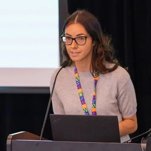
Portrait de Julien Cohen-Adad

Julien Cohen-Adad

Membre académique associé
Professeur agrégé, Polytechnique Montréal, Département de génie électrique
Professeur asssocié, Université de Montréal, Département de neurosciences
Sujets de recherche
Apprentissage automatique médical

Biographie

Julien Cohen-Adad est professeur à Polytechnique Montréal et directeur associé de l'Unité de neuro-imagerie fonctionnelle de l'Université de Montréal. Il est également titulaire de la Chaire de recherche du Canada en imagerie par résonance magnétique quantitative. Ses recherches portent sur l'avancement des méthodes de neuro-imagerie avec l'aide de l'IA. Voici quelques exemples de ses projets :

- Formation multimodale pour les tâches d'imagerie médicale (segmentation des pathologies, diagnostic, etc.);

- Ajout d'un a priori issu de la physique de l'IRM pour améliorer la généralisation des modèles;

- Incorporation de mesures d'incertitude pour traiter la variabilité interévaluateurs;

- Stratégies d'apprentissage continu lorsque le partage des données est restreint;

- Introduction des méthodes d'IA dans la routine de la radiologie clinique par l’intermédiaire de solutions logicielles conviviales.

Le professeur Cohen-Adad dirige également de nombreux projets de logiciels libres qui profitent à la communauté scientifique et clinique. Plus de détails sur https://neuro.polymtl.ca/software.html.

En résumé, Julien aime : l'IRM avec des aimants puissants, la neuro-imagerie, la programmation et la science ouverte!

Étudiants actuels

Stagiaire de recherche - Polytechnique
Doctorat - Polytechnique
Co-superviseur⋅e :
Doctorat - Polytechnique
Maîtrise recherche - Polytechnique
Doctorat - Polytechnique
Co-superviseur⋅e :
Maîtrise recherche - Polytechnique
Doctorat - Polytechnique
Collaborateur·rice de recherche
Maîtrise recherche - Polytechnique
Postdoctorat - Polytechnique

Publications

Titre: Title: Comparison of Myelin Imaging Techniques in Ex Vivo Spinal Cord Auteur:
Nikola Stikov
Manh-Tung Vuong
Vuong Manh Tung
Myelin is a dielectric material that wraps around the axons of nerve fibers to enable fast conduction of signals throughout the nervous syst… (voir plus)em. Loss of myelin can cause anywhere from minor interruption to complete disruption of nerve impulses in a range of neurodegenerative diseases such as multiple sclerosis and Parkinson’s disease. There is an ongoing debate in the myelin imaging community about which biomarker based on Magnetic Resonance Imaging (MRI) is more correlated with myelin. In this work, we implemented and compared several MRI-based myelin imaging techniques (quantitative magnetization transfer imaging, myelin water imaging, and proton density imaging) by evaluating their repeatability and their relation to large-scale histology in the ex vivo spinal cords of a rat, a dog, and a human. While there are studies investigating the relationship between pairs of them as well as with histology, to the best of our knowledge, this is the first study that implemented and compared all those methods at the same time to evaluate their reproducibility and their correlation with myelin. Qualitatively the contrasts were similar, and all techniques had comparable scan-rescan and correlations with histology. Surprisingly, the voxel-wise correlations between the various myelin measures were almost as high as the scan-rescan correlations. The correlations decreased when only white matter was considered, which could be due to the small dynamic range of the measurement, or due to artifacts related to the preparation and panoramic scanning of the tissue. We conclude that the myelin imaging techniques explored in this thesis exhibit similar specificity to myelin, yet the histological correlations suggest that more work is needed to determine the optimal myelin imaging protocol. The study also pointed out some potential miscalibrations during acquisitions as well as data processing that may lead to anywhere from minor to major impact on the accuracy of the results. These include B1 mapping, insufficient spoiling and variation of the predelay time. We have also standardized the data processing routines by upgrading qMTLab to qMRLab which adds several quantitative MR methods to the toolbox, such as standard T1 mapping and field mapping. In addition, the data of the dog spinal cord in this study will be published together with the analysis scripts to help the interested reader to reproduce the findings from this thesis.
The Myelin‐Weighted Connectome in Parkinson's Disease
Tommy Boshkovski
Bratislav Mišić
Isabelle Arnulf
Jean‐Christophe Corvol
Marie Vidailhet
Stéphane Lehéricy
Nikola Stikov
Matteo Mancini
2D Multi-Class Model for Gray and White Matter Segmentation of the Cervical Spinal Cord at 7T
Charley Gros
Virginie Callot
Arnaud Le Troter
Minimum detectable spinal cord atrophy with automatic segmentation: Investigations using an open-access dataset of healthy participants
Paul Bautin
Normalizing automatic spinal cord cross-sectional area measures
S. Bédard
Spinal cord cross-sectional area (CSA) is a relevant biomarker to assess spinal cord atrophy in various neurodegenerative diseases. However,… (voir plus) the considerable inter-subject variability among healthy participants currently limits its usage. Previous studies explored factors contributing to the variability, yet the normalization models were based on a relatively limited number of participants (typically 300 participants), required manual intervention, and were not implemented in an open-access comprehensive analysis pipeline. Another limitation is related to the imprecise prediction of the spinal levels when using vertebral levels as a reference; a question never addressed before in the search for a normalization method. In this study we implemented a method to measure CSA automatically from a spatial reference based on the central nervous system (the pontomedullary junction, PMJ), we investigated various factors to explain variability, and we developed normalization strategies on a large cohort (N=804). Cervical spinal cord CSA was computed on T1w MRI scans for 804 participants from the UK Biobank database. In addition to computing cross-sectional at the C2-C3 vertebral disc, it was also measured at 64 mm caudal from the PMJ. The effect of various biological, demographic and anatomical factors was explored by computing Pearson’s correlation coefficients. A stepwise linear regression found significant predictors; the coefficients of the best fit model were used to normalize CSA. The correlation between CSA measured at C2-C3 and using the PMJ was y = 0.98x + 1.78 (R2 = 0.97). The best normalization model included thalamus volume, brain volume, sex and interaction between brain volume and sex. With this model, the coefficient of variation went down from 10.09% (without normalization) to 8.59%, a reduction of 14.85%. In this study we identified factors explaining inter-subject variability of spinal cord CSA over a large cohort of participants, and developed a normalization model to reduce the variability. We implemented an approach, based on the PMJ, to measure CSA to overcome limitations associated with the vertebral reference. This approach warrants further validation, especially in longitudinal cohorts. The PMJ-based method and normalization models are readily available in the Spinal Cord Toolbox.
Author Correction: Open-access quantitative MRI data of the spinal cord and reproducibility across participants, sites and manufacturers
Eva Alonso‐Ortiz
Mihael Abramovic
Carina Arneitz
Nicole Atcheson
Laura Barlow
Robert L. Barry
Markus Barth
Marco Battiston
Christian Büchel
Matthew D. Budde
Virginie Callot
Anna J. E. Combes
Benjamin De Leener
Maxime Descoteaux
Paulo Loureiro de Sousa
Marek Dostál
Julien Doyon
Adam Dvorak
Falk Eippert … (voir 71 de plus)
Karla R. Epperson
Kevin S. Epperson
Patrick Freund
Jürgen Finsterbusch
Alexandru Foias
Michela Fratini
Issei Fukunaga
Claudia A. M. Gandini Wheeler-Kingshott
Giancarlo Germani
Guillaume Gilbert
Federico Giove
Charley Gros
Francesco Grussu
Akifumi Hagiwara
Pierre-Gilles Henry
Tomáš Horák
Masaaki Hori
James Joers
Kouhei Kamiya
Haleh Karbasforoushan
Miloš Keřkovský
Ali Khatibi
Joo‐Won Kim
Nawal Kinany
Hagen H. Kitzler
Shannon Kolind
Yazhuo Kong
Petr Kudlička
Paul Kuntke
Nyoman D. Kurniawan
Slawomir Kusmia
René Labounek
Maria Marcella Lagana
Cornelia Laule
Christine S. Law
Christophe Lenglet
Tobias Leutritz
Yaou Liu
Sara Llufriu
Sean Mackey
Eloy Martinez-Heras
Loan Mattera
Igor Nestrašil
Kristin P. O’Grady
Nico Papinutto
Daniel Papp
Deborah Pareto
Todd B. Parrish
Anna Pichiecchio
Ferran Prados
Àlex Rovira
Marc J. Ruitenberg
Rebecca S. Samson
Giovanni Savini
Maryam Seif
Alan C. Seifert
Alex K. Smith
Seth A. Smith
Zachary A. Smith
Elisabeth Solana
Y. Suzuki
George Tackley
Alexandra Tinnermann
Dimitri Van De Ville
Marios C. Yiannakas
Kenneth A. Weber
Nikolaus Weiskopf
Richard G. Wise
Patrik O. Wyss
Junqian Xu
Stacked Hourglass Network with a Multi-level Attention Mechanism: Where to Look for Intervertebral Disc Labeling
Reza Azad
Lucas Rouhier
Team NeuroPoly: Description of the Pipelines for the MICCAI 2021 MS New Lesions Segmentation Challenge
Enamundram Naga Karthik
Charley Gros
This paper gives a detailed description of the pipelines used for the 2nd edition of the MICCAI 2021 Challenge on Multiple Sclerosis Lesion … (voir plus)Segmentation. An overview of the data preprocessing steps applied is provided along with a brief description of the pipelines used, in terms of the architecture and the hyperparameters. Our code for this work can be found at: https://github.com/ivadomed/ms-challenge-2021.
Rapid simultaneous acquisition of macromolecular tissue volume, susceptibility, and relaxometry maps
Fang Frank Yu
Susie Yi Huang
Thomas Witzel
Ashwin Kumar
Congyu Liao
Tanguy Duval
Berkin Bilgic
Purpose A major obstacle to the clinical implementation of quantitative MR is the lengthy acquisition time required to derive multi-contrast… (voir plus) parametric maps. We sought to reduce the acquisition time for quantitative susceptibility mapping (QSM) and macromolecular tissue volume (MTV) by acquiring both contrasts simultaneously by leveraging their redundancies. The Joint Virtual Coil concept with generalized autocalibrating partially parallel acquisitions (JVC-GRAPPA) was applied to reduce acquisition time further. Methods Three adult volunteers were imaged on a 3T scanner using a multi-echo 3D GRE sequence acquired at three head orientations. MTV, QSM, R2*, T1, and proton density maps were reconstructed. The same sequence (GRAPPA R=4) was performed in subject #1 with a single head orientation for comparison. Fully sampled data was acquired in subject #2, from which retrospective undersampling was performed (R=6 GRAPPA and R=9 JVC-GRAPPA). Prospective undersampling was performed in subject #3 (R=6 GRAPPA and R=9 JVC-GRAPPA) using gradient blips to shift k-space sampling in later echoes. Results Subject #1’s multi-orientation and single-orientation MTV maps were not significantly different based on RMSE. For subject #2, the retrospectively undersampled JVC-GRAPPA and GRAPPA generated similar results as fully sampled data. This approach was validated with the prospectively undersampled images in subject #3. Using QSM, R2*, and MTV, the contributions of myelin and iron content to susceptibility was estimated. Conclusion We have developed a novel strategy to simultaneously acquire data for the reconstruction of five intrinsically co-registered 1-mm isotropic resolution multi-parametric maps, with a scan time of 6 minutes using JVC-GRAPPA.
Quantitative 7-Tesla Imaging of Cortical Myelin Changes in Early Multiple Sclerosis
Valeria Barletta
Elena Herranz
Constantina A. Treaba
Ambica Mehndiratta
Russell Ouellette
Gabriel Mangeat
Tobias Granberg
Jacob A. Sloane
Eric C Klawiter
Caterina Mainero
Cortical demyelination occurs early in multiple sclerosis (MS) and relates to disease outcome. The brain cortex has endogenous propensity fo… (voir plus)r remyelination as proven from histopathology study. In this study, we aimed at characterizing cortical microstructural abnormalities related to myelin content by applying a novel quantitative MRI technique in early MS. A combined myelin estimation (CME) cortical map was obtained from quantitative 7-Tesla (7T) T2* and T1 acquisitions in 25 patients with early MS and 19 healthy volunteers. Cortical lesions in MS patients were classified based on their myelin content by comparison with CME values in healthy controls as demyelinated, partially demyelinated, or non-demyelinated. At follow-up, we registered changes in cortical lesions as increased, decreased, or stable CME. Vertex-wise analysis compared cortical CME in the normal-appearing cortex in 25 MS patients vs. 19 healthy controls at baseline and investigated longitudinal changes at 1 year in 10 MS patients. Measurements from the neurite orientation dispersion and density imaging (NODDI) diffusion model were obtained to account for cortical neurite/dendrite loss at baseline and follow-up. Finally, CME maps were correlated with clinical metrics. CME was overall low in cortical lesions (p = 0.03) and several normal-appearing cortical areas (p 0.05) in the absence of NODDI abnormalities. Individual cortical lesion analysis revealed, however, heterogeneous CME patterns from extensive to partial or absent demyelination. At follow-up, CME overall decreased in cortical lesions and non-lesioned cortex, with few areas showing an increase (p 0.05). Cortical CME maps correlated with processing speed in several areas across the cortex. In conclusion, CME allows detection of cortical microstructural changes related to coexisting demyelination and remyelination since the early phases of MS, and shows to be more sensitive than NODDI and relates to cognitive performance.
Generic acquisition protocol for quantitative MRI of the spinal cord
Eva Alonso‐Ortiz
Mihael Abramovic
Carina Arneitz
Nicole Atcheson
Laura Barlow
Robert L. Barry
Markus Barth
Marco Battiston
Christian Büchel
Matthew D. Budde
Virginie Callot
Anna J. E. Combes
Benjamin De Leener
Maxime Descoteaux
Paulo Loureiro de Sousa
Marek Dostál
Julien Doyon
Adam Dvorak
Falk Eippert … (voir 71 de plus)
Karla R. Epperson
Kevin S. Epperson
Patrick Freund
Jürgen Finsterbusch
Alexandru Foias
Michela Fratini
Issei Fukunaga
Claudia A. M. Gandini Wheeler-Kingshott
Giancarlo Germani
Guillaume Gilbert
Federico Giove
Charley Gros
Francesco Grussu
Akifumi Hagiwara
Pierre-Gilles Henry
Tomáš Horák
Masaaki Hori
James Joers
Kouhei Kamiya
Haleh Karbasforoushan
Miloš Keřkovský
Ali Khatibi
Joo‐Won Kim
Nawal Kinany
Hagen H. Kitzler
Shannon Kolind
Yazhuo Kong
Petr Kudlička
Paul Kuntke
Nyoman D. Kurniawan
Slawomir Kusmia
René Labounek
Maria Marcella Lagana
Cornelia Laule
Christine S. Law
Christophe Lenglet
Tobias Leutritz
Yaou Liu
Sara Llufriu
Sean Mackey
Eloy Martinez-Heras
Loan Mattera
Igor Nestrašil
Kristin P. O’Grady
Nico Papinutto
Daniel Papp
Deborah Pareto
Todd B. Parrish
Anna Pichiecchio
Ferran Prados
Àlex Rovira
Marc J. Ruitenberg
Rebecca S. Samson
Giovanni Savini
Maryam Seif
Alan C. Seifert
Alex K. Smith
Seth A. Smith
Zachary A. Smith
Elisabeth Solana
Yuichi Suzuki
George Tackley
Alexandra Tinnermann
Dimitri Van De Ville
Marios C. Yiannakas
K. Weber
Nikolaus Weiskopf
Richard G. Wise
Patrik O. Wyss
Junqian Xu
Open-access quantitative MRI data of the spinal cord and reproducibility across participants, sites and manufacturers
Eva Alonso‐Ortiz
Mihael Abramovic
Carina Arneitz
Nicole Atcheson
Laura Barlow
Robert L. Barry
Markus Barth
Marco Battiston
Christian Büchel
Matthew D. Budde
Virginie Callot
Anna J. E. Combes
Benjamin De Leener
Maxime Descoteaux
Paulo Loureiro de Sousa
Marek Dostál
Julien Doyon
Adam Dvorak
Falk Eippert … (voir 71 de plus)
Karla R. Epperson
Kevin S. Epperson
Patrick Freund
Jürgen Finsterbusch
Alexandru Foias
Michela Fratini
Issei Fukunaga
Claudia A. M. Gandini Wheeler-Kingshott
Giancarlo Germani
Guillaume Gilbert
Federico Giove
Charley Gros
Francesco Grussu
Akifumi Hagiwara
Pierre-Gilles Henry
Tomáš Horák
Masaaki Hori
James Joers
Kouhei Kamiya
Haleh Karbasforoushan
Miloš Keřkovský
Ali Khatibi
Joo‐Won Kim
Nawal Kinany
Hagen H. Kitzler
Shannon Kolind
Yazhuo Kong
Petr Kudlička
Paul Kuntke
Nyoman D. Kurniawan
Slawomir Kusmia
René Labounek
Maria Marcella Lagana
Cornelia Laule
Christine S. Law
Christophe Lenglet
Tobias Leutritz
Yaou Liu
Sara Llufriu
Sean Mackey
Eloy Martinez-Heras
Loan Mattera
Igor Nestrašil
Kristin P. O’Grady
Nico Papinutto
Daniel Papp
Deborah Pareto
Todd B. Parrish
Anna Pichiecchio
Ferran Prados
Àlex Rovira
Marc J. Ruitenberg
Rebecca S. Samson
Giovanni Savini
Maryam Seif
Alan C. Seifert
Alex K. Smith
Seth A. Smith
Zachary A. Smith
Elisabeth Solana
Y. Suzuki
George Tackley
Alexandra Tinnermann
Dimitri Van De Ville
Marios C. Yiannakas
Kenneth A. Weber
Nikolaus Weiskopf
Richard G. Wise
Patrik O. Wyss
Junqian Xu