Mila organise son premier hackathon en informatique quantique le 21 novembre. Une journée unique pour explorer le prototypage quantique et l’IA, collaborer sur les plateformes de Quandela et IBM, et apprendre, échanger et réseauter dans un environnement stimulant au cœur de l’écosystème québécois en IA et en quantique.
Une nouvelle initiative pour renforcer les liens entre la communauté de recherche, les partenaires et les expert·e·s en IA à travers le Québec et le Canada, grâce à des rencontres et événements en présentiel axés sur l’adoption de l’IA dans l’industrie.
Nous utilisons des témoins pour analyser le trafic et l’utilisation de notre site web, afin de personnaliser votre expérience. Vous pouvez désactiver ces technologies à tout moment, mais cela peut restreindre certaines fonctionnalités du site. Consultez notre Politique de protection de la vie privée pour en savoir plus.
Paramètre des cookies
Vous pouvez activer et désactiver les types de cookies que vous souhaitez accepter. Cependant certains choix que vous ferez pourraient affecter les services proposés sur nos sites (ex : suggestions, annonces personnalisées, etc.).
Cookies essentiels
Ces cookies sont nécessaires au fonctionnement du site et ne peuvent être désactivés. (Toujours actif)
Cookies analyse
Acceptez-vous l'utilisation de cookies pour mesurer l'audience de nos sites ?
Multimedia Player
Acceptez-vous l'utilisation de cookies pour afficher et vous permettre de regarder les contenus vidéo hébergés par nos partenaires (YouTube, etc.) ?
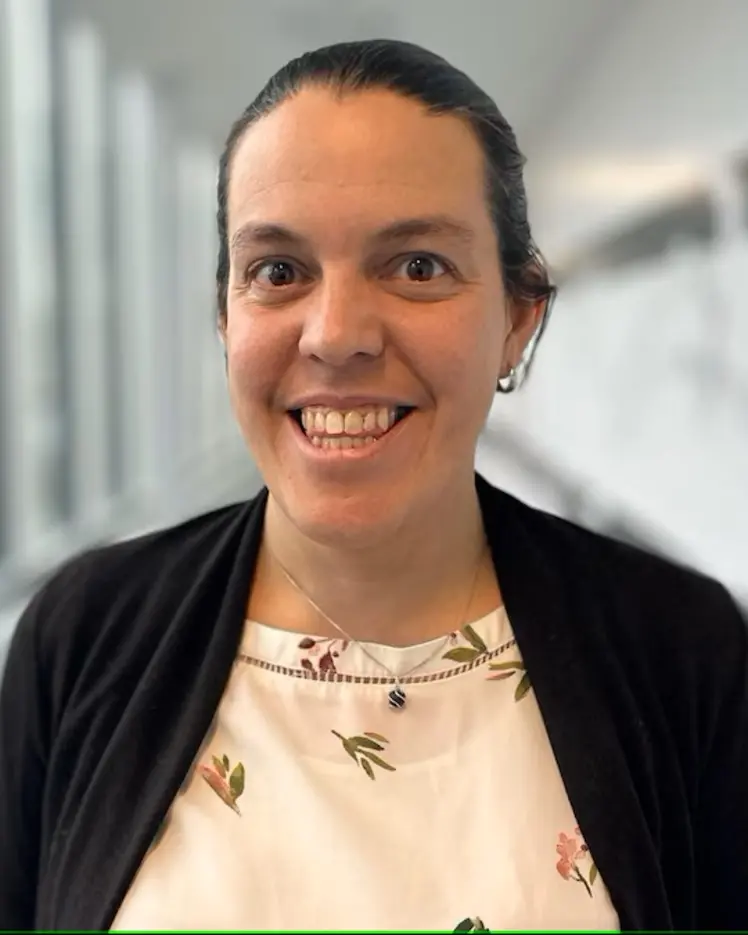
Flavie Lavoie-Cardinal
Membre académique associé
Professeur agrégé, Université Laval, Département de Psychiatrie et de neurosciences
The integration of artificial intelligence into microscopy systems significantly enhances performance, optimizing both the image acquisition… (voir plus) and analysis phases. Development of artificial intelligence (AI)-assisted super-resolution microscopy is often limited by the access to large biological datasets, as well as by the difficulties to benchmark and compare approaches on heterogeneous samples. We demonstrate the benefits of a realistic STED simulation platform, pySTED, for the development and deployment of AI-strategies for super-resolution microscopy. The simulation environment provided by pySTED allows the augmentation of data for the training of deep neural networks, the development of online optimization strategies, and the training of reinforcement learning models, that can be deployed successfully on a real microscope.
The integration of artificial intelligence (AI) into microscopy systems significantly enhances performance, optimizing both the image acquis… (voir plus)ition and analysis phases. Development of AI-assisted super-resolution microscopy is often limited by the access to large biological datasets, as well as by the difficulties to benchmark and compare approaches on heterogeneous samples. We demonstrate the benefits of a realistic STED simulation platform, pySTED, for the development and deployment of AI-strategies for super-resolution microscopy. The simulation environment provided by pySTED allows the augmentation of data for the training of deep neural networks, the development of online optimization strategies, and the training of reinforcement learning models, that can be deployed successfully on a real microscope.
Ca2+ imaging methods are widely used for studying cellular activity in the brain, allowing detailed analysis of dynamic processes across var… (voir plus)ious scales. Enhanced by high-contrast optical microscopy and fluorescent Ca2+ sensors, this technique can be used to reveal localized Ca2+ fluctuations within neurons, including in sub-cellular compartments, such as the dendritic shaft or spines. Despite advances in Ca2+ sensors, the analysis of miniature Synaptic Calcium Transients (mSCTs), characterized by variability in morphology and low signal-to-noise ratios, remains challenging. Traditional threshold-based methods struggle with the detection and segmentation of these small, dynamic events. Deep learning (DL) approaches offer promising solutions but are limited by the need for large annotated datasets. Positive Unlabeled (PU) learning addresses this limitation by leveraging unlabeled instances to increase dataset size and enhance performance. This approach is particularly useful in the case of mSCTs that are scarce and small, associated with a very small proportion of the foreground pixels. PU learning significantly increases the effective size of the training dataset, improving model performance. Here, we present a PU learning-based strategy for detecting and segmenting mSCTs. We evaluate the performance of two 3D deep learning models, StarDist-3D and 3D U-Net, which are well established for the segmentation of small volumetric structures in microscopy datasets. By integrating PU learning, we enhance the 3D U-Net’s performance, demonstrating significant gains over traditional methods. This work pioneers the application of PU learning in Ca2+ imaging analysis, offering a robust framework for mSCT detection and segmentation. We also demonstrate how this quantitative analysis pipeline can be used for subsequent mSCTs feature analysis. We characterize morphological and kinetic changes of mSCTs associated with the application of chemical long-term potentiation (cLTP) stimulation in cultured rat hippocampal neurons. Our data-driven approach shows that a cLTP-inducing stimulus leads to the emergence of new active dendritic regions and differently affects mSCTs subtypes.
The development of signal unmixing algorithms is essential for leveraging multimodal datasets acquired through a wide array of scientific im… (voir plus)aging technologies, including hyperspectral or time-resolved acquisitions. In experimental physics, enhancing the spatio-temporal resolution or expanding the number of detection channels often leads to diminished sampling rate and signal-to-noise ratio (SNR), significantly affecting the efficacy of signal unmixing algorithms. We propose Latent Unmixing, a new approach which applies band-pass filters to the latent space of a multi-dimensional convolutional neural network to disentangle overlapping signal components. It enables better isolation and quantification of individual signal contributions, especially in the context of undersampled distributions. Using multi-dimensional convolution kernels to process all dimensions simultaneously enhances the network's ability to extract information from adjacent pixels, and time- or spectral-bins. This approach enables more effective separation of components in cases where individual pixels do not provide clear, well-resolved information. We showcase the method's practical use in experimental physics through two test cases that highlight the versatility of our approach: fluorescence lifetime microscopy and mode decomposition in optical fibers. The latent unmixing method extracts valuable information from complex signals that cannot be resolved by standard methods. It opens new possibilities in optics and photonics for multichannel separations at an increased sampling rate.