

Portrait of Danilo Bzdok

Danilo Bzdok

Core Academic Member
Canada CIFAR AI Chair
Associate Professor, McGill University, Department of Biomedical Engineering
Research Topics
Computational Biology
Deep Learning
Large Language Models (LLM)
Natural Language Processing

Biography

Danilo Bzdok is a computer scientist and medical doctor by training with a unique dual background in systems neuroscience and machine learning algorithms. After training at RWTH Aachen University (Germany), Université de Lausanne (Switzerland) and Harvard Medical School, Bzdok completed two doctoral degrees, one in neuroscience at Forschungszentrum Jülich in Germany, and another in computer science (machine learning statistics) at INRIA–Saclay and the Neurospin brain imaging centre in Paris.

Danilo is currently an associate professor at McGill University’s Faculty of Medicine and a Canada CIFAR AI Chair at Mila – Quebec Artificial Intelligence Institute. His interdisciplinary research centres around narrowing knowledge gaps in the brain basis of human-defining types of thinking in order to uncover key computational design principles underlying human intelligence.
Current Students

PhD - McGill University
Undergraduate - CentraleSupélec
PhD - McGill University
Collaborating researcher - École Polytechnique Montréal Paris
PhD - McGill University
Postdoctorate - McGill University
Master's Research - McGill University
Independent visiting researcher - McGill University
PhD - McGill University
Collaborating researcher - Aix-Marseille Université
PhD - McGill University

Publications

Systematic cross-sectional age-associations in global fMRI signal topography
Jason S. Nomi
Jingwei Li
Taylor Bolt
Catie Chang
Salome Kornfeld
Zachary T. Goodman
B.T. Thomas Yeo
R. Nathan Spreng
Lucina Q. Uddin
The global signal (GS) in resting-state functional MRI (fMRI), known to contain artifacts and non-neuronal physiological signals, also conta… (see more)ins important neural information related to individual state and trait characteristics. Here, we show distinct linear and curvilinear relationships between GS topography and age in a cross-sectional sample of individuals (6-85 years old) representing a significant portion of the lifespan. Subcortical brain regions such as the thalamus and putamen show linear associations with the GS across age. The thalamus has stronger contributions to the GS in older-age individuals compared with younger-aged individuals, while the putamen has stronger contributions in younger individuals compared with older individuals. The subcortical nucleus basalis of Meynert shows a u-shaped pattern similar to cortical regions within the lateral frontoparietal network and dorsal attention network, where contributions of the GS are stronger at early and old age, and weaker in middle age. This differentiation between subcortical and cortical brain activity across age supports a dual-layer model of GS composition, where subcortical aspects of the GS are differentiated from cortical aspects of the GS. We find that these subcortical-cortical contributions to the GS depend strongly on age across the lifespan of human development. Our findings demonstrate how neurobiological information within the GS differs across development and highlight the need to carefully consider whether or not to remove this signal when investigating age-related functional differences in the brain.
Harnessing population diversity: in search of tools of the trade
Big neuroscience datasets are not big small datasets when it comes to quantitative data analysis. Neuroscience has now witnessed the advent … (see more)of many population cohort studies that deep-profile participants, yielding hundreds of measures, capturing dimensions of each individual’s position in the broader society. Indeed, there is a rebalancing from small, strictly selected, and thus homogenized cohorts toward always larger, more representative, and thus diverse cohorts. This shift in cohort composition is prompting the revision of incumbent modeling practices. Major sources of population stratification increasingly overshadow the subtle effects that neuroscientists are typically studying. In our opinion, as we sample individuals from always wider diversity backgrounds, we will require a new stack of quantitative tools to realize diversity-aware modeling. We here take inventory of candidate analytical frameworks. Better incorporating driving factors behind population structure will allow refining our understanding of how brain–behavior relationships depend on human subgroups.
Harnessing population diversity: in search of tools of the trade
Abstract Big neuroscience datasets are not big small datasets when it comes to quantitative data analysis. Neuroscience has now witnessed th… (see more)e advent of many population cohort studies that deep-profile participants, yielding hundreds of measures, capturing dimensions of each individual’s position in the broader society. Indeed, there is a rebalancing from small, strictly selected, and thus homogenized cohorts toward always larger, more representative, and thus diverse cohorts. This shift in cohort composition is prompting the revision of incumbent modeling practices. Major sources of population stratification increasingly overshadow the subtle effects that neuroscientists are typically studying. In our opinion, as we sample individuals from always wider diversity backgrounds, we will require a new stack of quantitative tools to realize diversity-aware modeling. We here take inventory of candidate analytical frameworks. Better incorporating driving factors behind population structure will allow refining our understanding of how brain–behavior relationships depend on human subgroups.
ImageFlowNet: Forecasting Multiscale Image-Level Trajectories of Disease Progression with Irregularly-Sampled Longitudinal Medical Images
Chen Liu
Ke Xu
Liangbo L. Shen
Jay Stewart
Jay C. Wang
Lucian V. Del Priore
Advances in medical imaging technologies have enabled the collection of longitudinal images, which involve repeated scanning of the same pat… (see more)ients over time, to monitor disease progression. However, predictive modeling of such data remains challenging due to high dimensionality, irregular sampling, and data sparsity. To address these issues, we propose ImageFlowNet, a novel model designed to forecast disease trajectories from initial images while preserving spatial details. ImageFlowNet first learns multiscale joint representation spaces across patients and time points, then optimizes deterministic or stochastic flow fields within these spaces using a position-parameterized neural ODE/SDE framework. The model leverages a UNet architecture to create robust multiscale representations and mitigates data scarcity by combining knowledge from all patients. We provide theoretical insights that support our formulation of ODEs, and motivate our regularizations involving high-level visual features, latent space organization, and trajectory smoothness. We validate ImageFlowNet on three longitudinal medical image datasets depicting progression in geographic atrophy, multiple sclerosis, and glioblastoma, demonstrating its ability to effectively forecast disease progression and outperform existing methods. Our contributions include the development of ImageFlowNet, its theoretical underpinnings, and empirical validation on real-world datasets. The official implementation is available at https://github.com/KrishnaswamyLab/ImageFlowNet.
ImageFlowNet: Forecasting Multiscale Image-Level Trajectories of Disease Progression with Irregularly-Sampled Longitudinal Medical Images
Chen Liu
Ke Xu
Liangbo L. Shen
Jay Stewart
Jay C. Wang
Lucian V. Del Priore
Advances in medical imaging technologies have enabled the collection of longitudinal images, which involve repeated scanning of the same pat… (see more)ients over time, to monitor disease progression. However, predictive modeling of such data remains challenging due to high dimensionality, irregular sampling, and data sparsity. To address these issues, we propose ImageFlowNet, a novel model designed to forecast disease trajectories from initial images while preserving spatial details. ImageFlowNet first learns multiscale joint representation spaces across patients and time points, then optimizes deterministic or stochastic flow fields within these spaces using a position-parameterized neural ODE/SDE framework. The model leverages a UNet architecture to create robust multiscale representations and mitigates data scarcity by combining knowledge from all patients. We provide theoretical insights that support our formulation of ODEs, and motivate our regularizations involving high-level visual features, latent space organization, and trajectory smoothness. We validate ImageFlowNet on three longitudinal medical image datasets depicting progression in geographic atrophy, multiple sclerosis, and glioblastoma, demonstrating its ability to effectively forecast disease progression and outperform existing methods. Our contributions include the development of ImageFlowNet, its theoretical underpinnings, and empirical validation on real-world datasets. The official implementation is available at https://github.com/KrishnaswamyLab/ImageFlowNet.
Performance reserves in brain-imaging-based phenotype prediction
Marc-Andre Schulz
Stefan Haufe
John-Dylan Haynes
Kerstin Ritter
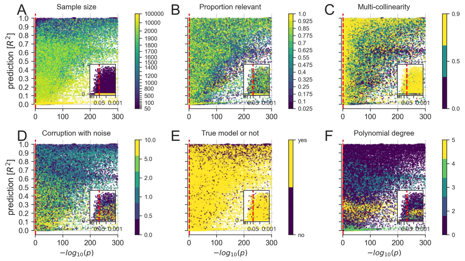
This study examines the impact of sample size on predicting cognitive and mental health phenotypes from brain imaging via machine learning. … (see more)Our analysis shows a 3- to 9-fold improvement in prediction performance when sample size increases from 1,000 to 1 M participants. However, despite this increase, the data suggest that prediction accuracy remains worryingly low and far from fully exploiting the predictive potential of brain imaging data. Additionally, we find that integrating multiple imaging modalities boosts prediction accuracy, often equivalent to doubling the sample size. Interestingly, the most informative imaging modality often varied with increasing sample size, emphasizing the need to consider multiple modalities. Despite significant performance reserves for phenotype prediction, achieving substantial improvements may necessitate prohibitively large sample sizes, thus casting doubt on the practical or clinical utility of machine learning in some areas of neuroimaging.
Translating phenotypic prediction models from big to small anatomical MRI data using meta-matching
Naren Wulan
Lijun An
Chen Zhang
Ru Kong
Pansheng Chen
Simon B. Eickhoff
Avram J. Holmes
B.T. Thomas Yeo
Individualized phenotypic prediction based on structural MRI is an important goal in neuroscience. Prediction performance increases with lar… (see more)ger samples, but small-scale datasets with fewer than 200 participants are often unavoidable. We have previously proposed a “meta-matching” framework to translate models trained from large datasets to improve the prediction of new unseen phenotypes in small collection efforts. Meta-matching exploits correlations between phenotypes, yielding large improvement over classical machine learning when applied to prediction models using resting-state functional connectivity as input features. Here, we adapt the two best performing meta-matching variants (“meta-matching finetune” and “meta-matching stacking”) from our previous study to work with T1-weighted MRI data by changing the base neural network architecture to a 3D convolution neural network. We compare the two meta-matching variants with elastic net and classical transfer learning using the UK Biobank (N = 36,461), Human Connectome Project Young Adults (HCP-YA) dataset (N = 1,017) and HCP-Aging dataset (N = 656). We find that meta-matching outperforms elastic net and classical transfer learning by a large margin, both when translating models within the same dataset, as well as translating models across datasets with different MRI scanners, acquisition protocols and demographics. For example, when translating a UK Biobank model to 100 HCP-YA participants, meta-matching finetune yielded a 136% improvement in variance explained over transfer learning, with an average absolute gain of 2.6% (minimum = −0.9%, maximum = 17.6%) across 35 phenotypes. Overall, our results highlight the versatility of the meta-matching framework.
Dissociable influences of maternal vs paternal Alzheimer’s risk on neurocognitive and cardiovascular health in men and women
Frederic St‐Onge
Sylvia Villeneuve
AmanPreet Badhwar
Sarah A Gagliano Taliun
Sali Farhan
Maiya R. Geddes
Yasser Iturria Medina
Judes Poirier
R. Nathan Spreng
We uncovered IPs of AD susceptibility differently expressed in male and female probands and affected by the diagnosed parent’s sex. Matern… (see more)al inheritance highlighted memory performance in both sexes, whereas paternal inheritance was particularly linked to cardiovascular health in males. The inheritance of the IPs was reflected in the brain structure at both superficial and deeper layers of the cortex. As the first study of its kind, our cross‐generational analysis of matri‐ vs. patrilinear AD risk bridges the epidemiological and clinical literature by leveraging the power of ∼1,000 patient visits. Our completely data‐driven framework ultimately dissociated phenotypes of maternal and paternal AD risk single‐handedly expressed in male and female probands.
Simulation‐based effect size analysis in the absence of drug effects to inform the design of clinical trials in Alzheimer’s disease
Daniel Andrews
Douglas L Arnold
Simon Ducharme
Howard Chertkow
D Louis Collins
Our results suggest low false positive probabilities for the successful trials. Cohort composition, sampling, and other trial characteristic… (see more)s might influence treatment‐independent group difference in cognitive decline rate. With our method, future trials could target treatment effects outside the target population’s false positive range.
General anaesthesia reduces the uniqueness of brain connectivity across individuals and across species
Andrea I. Luppi
Daniel Golkowski
Andreas Ranft
Rudiger Ilg
Denis Jordan
Adrian M. Owen
Lorina Naci
Emmanuel A. Stamatakis
Enrico Amico
Bratislav Misic
The human brain is characterised by idiosyncratic patterns of spontaneous thought, rendering each brain uniquely identifiable from its neura… (see more)l activity. However, deep general anaesthesia suppresses subjective experience. Does it also suppress what makes each brain unique? Here we used functional MRI under the effects of the general anaesthetics sevoflurane and propofol to determine whether anaesthetic-induced unconsciousness diminishes the uniqueness of the human brain: both with respect to the brains of other individuals, and the brains of another species. We report that under anaesthesia individual brains become less self-similar and less distinguishable from each other. Loss of distinctiveness is highly organised: it co-localises with the archetypal sensory-association axis, correlating with genetic and morphometric markers of phylogenetic differences between humans and other primates. This effect is more evident at greater anaesthetic depths, reproducible across sevoflurane and propofol, and reversed upon recovery. Providing convergent evidence, we show that under anaesthesia the functional connectivity of the human brain becomes more similar to the macaque brain. Finally, anaesthesia diminishes the match between spontaneous brain activity and meta-analytic brain patterns aggregated from the NeuroSynth engine. Collectively, the present results reveal that anaesthetised human brains are not only less distinguishable from each other, but also less distinguishable from the brains of other primates, with specifically human-expanded regions being the most affected by anaesthesia.
Aberrant functional brain network organization is associated with relapse during 1-year follow-up in alcohol-dependent patients
Justin Böhmer
Pablo Reinhardt
Maria Garbusow
Michael Marxen
Michael N. Smolka
U. Zimmermann
Andreas Heinz
Eva Friedel
Johann Kruschwitz
Henrik Walter
Alcohol dependence (AD) is a debilitating disease associated with high relapse rates even after long periods of abstinence. Thus, elucidatin… (see more)g neurobiological substrates of relapse risk is fundamental for the development of novel targeted interventions that could promote long-lasting abstinence. In the present study, we analyzed resting-state functional magnetic resonance imaging (rsfMRI) data from a sample of recently detoxified AD patients (n = 93) who were followed-up for 12 months after rsfMRI assessment. Specifically, we employed graph theoretic analyses to compare functional brain network topology and functional connectivity between future relapsers (REL, n = 59), future abstainers (ABS, n = 28) and age and gender matched controls (CON, n = 83). Our results suggest increased whole-brain network segregation, decreased global network integration and overall blunted connectivity strength in REL compared to CON. Conversely, we found evidence for a comparable network architecture in ABS relative to CON. At the nodal level, REL exhibited decreased integration and decoupling between multiple brain systems compared to CON, encompassing regions associated with higher-order executive functions, sensory and reward processing. Among AD patients, increased coupling between nodes implicated in reward valuation and salience attribution constitutes a particular risk factor for future relapse. Importantly, aberrant network organization in REL was consistently associated with shorter abstinence duration during follow-up, portending to a putative neural signature of relapse risk in AD. Future research should further evaluate the potential diagnostic value of the identified changes in network topology and functional connectivity for relapse prediction at the individual subject level.
Bayesian modelling disentangles language versus executive control disruption in stroke
Gesa Hartwigsen
Jae-Sung Lim
Hee-Joon Bae
Kyung-Ho Yu
Hugo J. Kuijf
Nick A. Weaver
J. Matthijs Biesbroek
Stroke is the leading cause of long-term disability worldwide. Incurred brain damage disrupts cognition, often with persisting deficits in l… (see more)anguage and executive capacities. Despite their clinical relevance, the commonalities, and differences of language versus executive control impairments remain under-specified. We tailored a Bayesian hierarchical modeling solution in a largest-of-its-kind cohort (1080 stroke patients) to deconvolve language and executive control in the brain substrates of stroke insults. Four cognitive factors distinguished left- and right-hemispheric contributions to ischemic tissue lesion. One factor delineated language and general cognitive performance and was mainly associated with damage to left-hemispheric brain regions in the frontal and temporal cortex. A factor for executive control summarized control and visual-constructional abilities. This factor was strongly related to right-hemispheric brain damage of posterior regions in the occipital cortex. The interplay of language and executive control was reflected in two factors: executive speech functions and verbal memory. Impairments on both were mainly linked to left-hemispheric lesions. These findings shed light onto the causal implications of hemispheric specialization for cognition; and make steps towards subgroup-specific treatment protocols after stroke.