

Portrait of Danilo Bzdok

Danilo Bzdok

Core Academic Member
Canada CIFAR AI Chair
Associate Professor, McGill University, Department of Biomedical Engineering
Research Topics
Computational Biology
Deep Learning
Large Language Models (LLM)
Natural Language Processing

Biography

Danilo Bzdok is a computer scientist and medical doctor by training with a unique dual background in systems neuroscience and machine learning algorithms. After training at RWTH Aachen University (Germany), Université de Lausanne (Switzerland) and Harvard Medical School, Bzdok completed two doctoral degrees, one in neuroscience at Forschungszentrum Jülich in Germany, and another in computer science (machine learning statistics) at INRIA–Saclay and the Neurospin brain imaging centre in Paris.

Danilo is currently an associate professor at McGill University’s Faculty of Medicine and a Canada CIFAR AI Chair at Mila – Quebec Artificial Intelligence Institute. His interdisciplinary research centres around narrowing knowledge gaps in the brain basis of human-defining types of thinking in order to uncover key computational design principles underlying human intelligence.
Current Students

PhD - McGill University
Undergraduate - CentraleSupélec
Collaborating researcher - École Polytechnique Montréal Paris
PhD - McGill University
Master's Research - McGill University
Independent visiting researcher - McGill University
PhD - McGill University
Collaborating researcher - Aix-Marseille Université
PhD - McGill University

Publications

Brain Diffusion Transformer for Personalized Neuroscience and Psychiatry
Rongquan Zhai
Yechen Hu
Liping Zheng
Shitong Xiang
Chao Xie
Lei Peng
Tobias Banaschewski
Gareth J. Barker
Arun L.W. Bokde
Rüdiger Brühl
Sylvane Desrivières
Herta Flor
Hugh Garavan
Penny Gowland
Antoine Grigis
Andreas Heinz
Herve Lemaitre
Jean-Luc Martinot
Marie-Laure Paillère Martinot
Eric Artiges … (see 26 more)
Frauke Nees
Dimitri Papadopoulos Orfanos
Luise Poustka
Michael N. Smolka
Sarah Hohmann
Nathalie Holz
Nilakshi Vaidya
Robert Whelan
Zuo Zhang
Lauren Robinson
Jeanne Winterer
Sinead King
Yuning Zhang
Hedi Kebir
Ulrike Schmidt
Julia Sinclair
Argyris Stringaris
Gunter Schumann
Henrik Walter
Edmund T. Rolls
Barbara Sahakian
Trevor W. Robbins
Jianfeng Feng
Weikang Gong
Tianye Jia
Task-fMRI analyses typically focus on localized activation contrasts between stimuli, neglecting the brain’s dynamic hierarchy. We introdu… (see more)ce Brain Diffusion Transformer (Brain-DiT), a deep generative model capturing recurrent processing underlying individualized neurocognitive state transitions via functional networks. Without prior assumptions, Brain-DiT identifies canonical cognitive regions in the brain and reveals replicable subgroups with distinct neural circuits in large cohorts, offering critical clinical insights overlooked by traditional methods: individuals exhibiting negative emotion bias, linked to language-related regions, had a 12-fold higher likelihood of major depression, and those with maladaptive inhibition strategies, associated with overactive medial frontal regions, showed a 9-fold increased risk of alcohol abuse. By bridging cognitive theory and psychiatric applications, Brain-DiT provides a unified analytical paradigm, paving the way for operational personalized medicine in psychiatry.
Steering CLIP's vision transformer with sparse autoencoders
Ethan Goldfarb
Lorenz Hufe
Yossi Gandelsman
Robert Graham
Wojciech Samek
Blake Aaron Richards
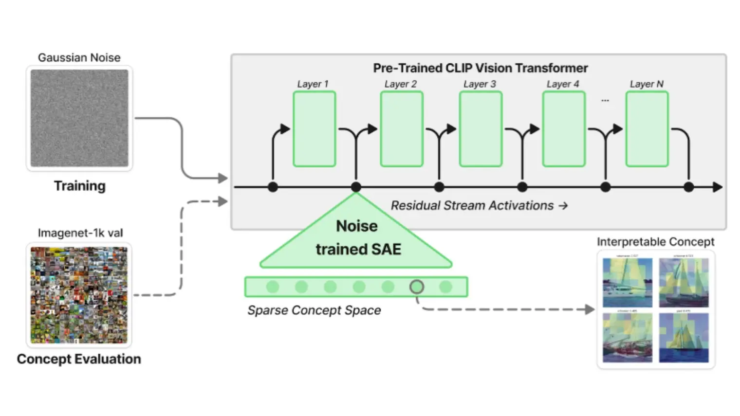
While vision models are highly capable, their internal mechanisms remain poorly understood-- a challenge which sparse autoencoders (SAEs) ha… (see more)ve helped address in language, but which remains underexplored in vision. We address this gap by training SAEs on CLIP's vision transformer and uncover key differences between vision and language processing, including distinct sparsity patterns for SAEs trained across layers and token types. We then provide the first systematic analysis of the steerability of CLIP's vision transformer by introducing metrics to quantify how precisely SAE features can be steered to affect the model's output. We find that 10-15% of neurons and features are steerable, with SAEs providing thousands more steerable features than the base model. Through targeted suppression of SAE features, we then demonstrate improved performance on three vision disentanglement tasks (CelebA, Waterbirds, and typographic attacks), finding optimal disentanglement in middle model layers, and achieving state-of-the-art performance on defense against typographic attacks. We release our CLIP SAE models and code to support future research in vision transformer interpretability.
General anaesthesia decreases the uniqueness of brain functional connectivity across individuals and species
Andrea I. Luppi
Daniel Golkowski
Andreas Ranft
Rudiger Ilg
Denis Jordan
Adrian M. Owen
Lorina Naci
Emmanuel A. Stamatakis
Enrico Amico
Bratislav Misic
The human brain is characterized by idiosyncratic patterns of spontaneous thought, rendering each brain uniquely identifiable from its neura… (see more)l activity. However, deep general anaesthesia suppresses subjective experience. Does it also suppress what makes each brain unique? Here we used functional MRI scans acquired under the effects of the general anaesthetics sevoflurane and propofol to determine whether anaesthetic-induced unconsciousness diminishes the uniqueness of the human brain, both with respect to the brains of other individuals and the brains of another species. Using functional connectivity, we report that under anaesthesia individual brains become less self-similar and less distinguishable from each other. Loss of distinctiveness is highly organized: it co-localizes with the archetypal sensory–association axis, correlating with genetic and morphometric markers of phylogenetic differences between humans and other primates. This effect is more evident at greater anaesthetic depths, reproducible across sevoflurane and propofol and reversed upon recovery. Providing convergent evidence, we show that anaesthesia shifts the functional connectivity of the human brain closer to the functional connectivity of the macaque brain in a low-dimensional space. Finally, anaesthesia diminishes the match between spontaneous brain activity and cognitive brain patterns aggregated from the Neurosynth meta-analytic engine. Collectively, the present results reveal that anaesthetized human brains are not only less distinguishable from each other, but also less distinguishable from the brains of other primates, with specifically human-expanded regions being the most affected by anaesthesia.
Quantifying associations between socio-spatial factors and cognitive development in the ABCD cohort
Quantifying associations between socio-spatial factors and cognitive development in the ABCD cohort.
Carriers of
<i>LRRK2</i>
pathogenic variants show a milder, anatomically distinct brain signature of Parkinson’s disease
Andrew Vo
Qin Tao
Tanya Simuni
Lana M. Chahine
Alain Dagher
LRRK2 gene variants are a major genetic risk factor for both familial and sporadic Parkinson’s disease (PD), opening an … (see more)unattended window on the disease’s mechanisms and potential therapies. Investigating the influence of pathogenic variants in LRRK2 gene on brain structure is a crucial step toward enabling early diagnosis and personalized treatment. Yet, despite its significance, the ways in which LRRK2 genotype affects brain structure remain largely unexplored. Work in this domain is plagued by small sample sizes and differences in cohort composition, which can obscure genuine distinctions among clinical subgroups. In this study, we overcome such important limitations by combining explicit modeling of population background variation and pattern matching. Specifically, we leveraged a large cohort of 641 participants (including 364 with a PD diagnosis) to examine MRI-detectable cortical atrophy patterns associated with the LRRK2 pathogenic variants in people with PD and non-manifesting individuals. LRRK2 PD patients exhibited milder cortical thinning compared to sporadic PD, with notable preservation in temporal and occipital regions, suggesting a distinct pattern of neurodegeneration. Non-manifesting LRRK2 carriers showed no significant cortical atrophy, indicating no structural signs of subclinical PD. We further analyzed the relationship between aggregated alpha-synuclein in cerebrospinal fluid and atrophy. We found that those with evidence of aggregated alpha-synuclein experienced pronounced neurodegeneration and increased cortical thinning, possibly defining another aggressive PD subtype. Our findings highlight avenues for distinguishing PD subtypes, which could lead to more targeted treatment approaches and a more complete understanding of Parkinson’s disease progression.
Large language models deconstruct the clinical intuition behind diagnosing autism
Emmett Rabot
Laurent Mottron
Cell type transcriptomics reveal shared genetic mechanisms in Alzheimer’s and Parkinson’s disease
Edward A. Fon
Alain Dagher
Yasser Iturria-Medina
Jo Anne Stratton
David A Bennett
Historically, Alzheimer’s disease (AD) and Parkinson’s disease (PD) have been investigated as two distinct disorders of the brain. Howev… (see more)er, a few similarities in neuropathology and clinical symptoms have been documented over the years. Traditional single gene-centric genetic studies, including GWAS and differential gene expression analyses, have struggled to unravel the molecular links between AD and PD. To address this, we tailor a pattern-learning framework to analyze synchronous gene co-expression at sub-cell-type resolution. Utilizing recently published single-nucleus AD (70,634 nuclei) and PD (340,902 nuclei) datasets from postmortem human brains, we systematically extract and juxtapose disease-critical gene modules. Our findings reveal extensive molecular similarities between AD and PD gene cliques. In neurons, disrupted cytoskeletal dynamics and mitochondrial stress highlight convergence in key processes; glial modules share roles in T-cell activation, myelin synthesis, and synapse pruning. This multi-module sub-cell-type approach offers insights into the molecular basis of shared neuropathology in AD and PD.
Cell type transcriptomics reveal shared genetic mechanisms in Alzheimer’s and Parkinson’s disease
Edward A. Fon
Alain Dagher
Yasser Iturria-Medina
Jo Anne Stratton
David A Bennett
Historically, Alzheimer’s disease (AD) and Parkinson’s disease (PD) have been investigated as two distinct disorders of the brain. Howev… (see more)er, a few similarities in neuropathology and clinical symptoms have been documented over the years. Traditional single gene-centric genetic studies, including GWAS and differential gene expression analyses, have struggled to unravel the molecular links between AD and PD. To address this, we tailor a pattern-learning framework to analyze synchronous gene co-expression at sub-cell-type resolution. Utilizing recently published single-nucleus AD (70,634 nuclei) and PD (340,902 nuclei) datasets from postmortem human brains, we systematically extract and juxtapose disease-critical gene modules. Our findings reveal extensive molecular similarities between AD and PD gene cliques. In neurons, disrupted cytoskeletal dynamics and mitochondrial stress highlight convergence in key processes; glial modules share roles in T-cell activation, myelin synthesis, and synapse pruning. This multi-module sub-cell-type approach offers insights into the molecular basis of shared neuropathology in AD and PD.
A hierarchical Bayesian brain parcellation framework for fusion of functional imaging datasets
Da Zhi
Caroline Nettekoven
Ana Lúısa Pinho
Jörn Diedrichsen
Inhibition of the inferior parietal lobe triggers state-dependent network adaptations
Kathleen A. Williams
Ole Numssen
Juan David Guerra
Gesa Hartwigsen
The human brain comprises large-scale networks that flexibly interact to support diverse cognitive functions and adapt to variability in dai… (see more)ly life. The inferior parietal lobe (IPL) is a hub of multiple brain networks that sustain various cognitive domains. It remains unclear how networks respond to acute regional perturbations to maintain normal function. To provoke network-level adaptive responses to local inhibition, we combined offline transcranial magnetic stimulation (TMS) over left or right IPL with neuroimaging during attention, semantic and social cognition tasks, and rest. Across tasks, TMS specifically affected task-active network activity with inhibition and facilitation. Network interaction responses differed between rest and tasks. After TMS over both IPL regions, large-scale network interactions were exclusively facilitated at rest, but mainly inhibited during tasks. Overall, responses to TMS primarily occurred in and between domain-general default mode and frontoparietal subnetworks. These findings elucidate short-term adaptive plasticity in response to network node inhibition.
Genetic Interplay Between White Matter Hyperintensities and Alzheimer's Disease: A Brain-Body Perspective
Manpreet Singh
Kimia Shafighi
Flavie E. Detcheverry
Fanta Dabo
Ikrame Housni
Sridar Narayanan
Sarah A. Gagliano Taliun
AmanPreet Badhwar
MRI-detected white matter hyperintensities (WMH) are often recognized as markers of cerebrovascular abnormalities and an index of vascular b… (see more)rain injury, and are frequently present in individuals with Alzheimer’s disease (AD). Given the emerging bidirectional communication between the brain-body axis in both WMHs and AD, it is important to understand their genetic underpinnings across the whole body. However, literature on this is scarce. We investigated the brain-body axis by breaking down heritability estimates of these phenotypes across the whole body, – i.e., partitioning heritability. Our aims were to identify genetic underpinnings specific to WMHs, and common between WMHs and AD, by assessing (a) the partitioned heritability of WMHs and AD across the brain-body axis with tissue-specific annotations, (b) the partitioned heritability of WMHs and AD across the brain-body axis with cell-specific annotations, and (c) the genes associated with WMHs and AD, and verifying their expression levels across the whole body. Our tissue-specific analysis revealed that WMH-associated SNPs were significantly enriched in tissues beyond the brain, namely liver, cardiovascular, and kidney – with liver being a common tissue enriched for both WMHs and AD. Our cell-specific analysis showed enrichment of vascular endothelial cells across the tissue types enriched for WMHs, highlighting their central role in the development of WMHs. Additionally, our gene-level analysis highlighted overlapping patterns of tissue enrichment for both WMHs and AD, and showed interactions between WMH and AD associated genes. Our findings provide new insights into the systemic influences potentially contributing to WMH pathology, in particular, multi-system endothelial disorder. We hope that our multisystemic genetic findings will stimulate future WMH-research into specific pathways across the brain-body axis.