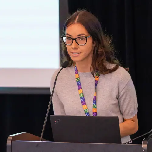
Portrait of Shirin A. Enger

Shirin A. Enger

Associate Academic Member
Tenured Associate Professor, McGill University, Department of Oncology
Research Topics
Computational Biology
Deep Learning
Medical Machine Learning

Biography

Shirin Abbasinejad Enger is a tenured associate professor in the Medical Physics Unit of the Gerald Bronfman Department of Oncology, McGill University.

She is also Director of the Medical Physics Unit, and a Tier 2 Canada Research Chair in Medical Physics.

Enger is also a principal investigator at the Lady Davis Institute for Medical Research and the Segal Cancer Centre of the Jewish General Hospital.

She received her PhD from Uppsala University in 2009 and was a postdoctoral fellow at Université Laval from 2009 to 2011. She has taken on a variety of leadership roles in international and national working groups and committees.

Current Students

Postdoctorate - McGill University
PhD - McGill University
Postdoctorate - McGill University
PhD - McGill University
Postdoctorate - McGill University
PhD - McGill University

Publications

Development of small, cost‐efficient scintillating fiber detectors for automated synthesis of positron emission tomography radiopharmaceuticals
Hailey Ahn
Liam Carroll
Robert Hopewell
I-Huang Tsai
Dean Jolly
Gassan Massarweh
Relative biological effectiveness of clinically relevant photon energies for the survival of human colorectal, cervical, and prostate cancer cell lines
Joanna Li
N. Chabaytah
Joud Babik
Behnaz Behmand
H. Bekerat
Tanner Connell
Michael D C Evans
Russell Ruo
T. Vuong
169Yb-based high dose rate intensity modulated brachytherapy for focal treatment of prostate cancer
Maude Robitaille
Cynthia Ménard
Gabriel Famulari
Dominic Béliveau-Nadeau
Pioneering women in nuclear and radiation sciences
Mirta Dumancic
Investigation of dosimetric characteristics of radiochromic film in response to alpha particles emitted from Americium-241.
Victor D. Diaz‐Martinez
Mélodie Cyr
S. Devic
Nada Tomic
David F. Lewis
BACKGROUND In radiotherapy, it is essential to deliver prescribed doses to tumors while minimizing damage to surrounding healthy tissue. Acc… (see more)urate measurements of absorbed dose are required for this purpose. Gafchromic® external beam therapy (EBT) radiochromic films have been widely used in radiotherapy. While the dosimetric characteristics of the EBT3 model film have been extensively studied for photon and charged particle beams (protons, electrons, and carbon ions), little research has been done on α
Automatic segmentation of Organs at Risk in Head and Neck cancer patients from CT and MRI scans
Sébastien Quetin
Andrew Heschl
Mauricio Murillo
Rohit Murali
Farhad Maleki
Background and purpose: Deep Learning (DL) has been widely explored for Organs at Risk (OARs) segmentation; however, most studies have focus… (see more)ed on a single modality, either CT or MRI, not both simultaneously. This study presents a high-performing DL pipeline for segmentation of 30 OARs from MRI and CT scans of Head and Neck (H&N) cancer patients. Materials and methods: Paired CT and MRI-T1 images from 42 H&N cancer patients alongside annotation for 30 OARs from the H&N OAR CT&MR segmentation challenge dataset were used to develop a segmentation pipeline. After cropping irrelevant regions, rigid followed by non-rigid registration of CT and MRI volumes was performed. Two versions of the CT volume, representing soft tissues and bone anatomy, were stacked with the MRI volume and used as input to an nnU-Net pipeline. Modality Dropout was used during the training to force the model to learn from the different modalities. Segmentation masks were predicted with the trained model for an independent set of 14 new patients. The mean Dice Score (DS) and Hausdorff Distance (HD) were calculated for each OAR across these patients to evaluate the pipeline. Results: This resulted in an overall mean DS and HD of 0.777 +- 0.118 and 3.455 +- 1.679, respectively, establishing the state-of-the-art (SOTA) for this challenge at the time of submission. Conclusion: The proposed pipeline achieved the best DS and HD among all participants of the H&N OAR CT and MR segmentation challenge and sets a new SOTA for automated segmentation of H&N OARs.
Characterizing the voxel-based approaches in radioembolization dosimetry with reDoseMC.
Taehyung Peter Kim
BACKGROUND Yttrium-90 ( 90 Y …
Penalty weight tuning in high dose rate brachytherapy using multi-objective Bayesian optimization.
Hossein Jafarzadeh
Majd Antaki
Ximeng Mao
Marie Duclos
Farhad Maleki
OBJECTIVE Treatment plan optimization in high dose rate (HDR) brachytherapy often requires manual fine-tuning of penalty weights for each ob… (see more)jective, which can be time-consuming and dependent on the planner's experience. To automate this process, this study used a multi-criteria approach called multi-objective Bayesian optimization with q-noisy expected hypervolume improvement as its acquisition function (MOBO-qNEHVI). Approach: The treatment plans of 13 prostate cancer patients were retrospectively imported to a research treatment planning system, RapidBrachyMTPS, where fast mixed integer optimization (FMIO) performs dwell time optimization given a set of penalty weights to deliver 15 Gy to the target volume. MOBO-qNEHVI was used to find patient-specific Pareto optimal penalty weight vectors that yield clinically acceptable dose volume histogram metrics. The relationship between the number of MOBO-qNEHVI iterations and the number of clinically acceptable plans per patient (acceptance rate) was investigated. The performance time was obtained for various parameter configurations. Main results: MOBO-qNEHVI found clinically acceptable treatment plans for all patients. With increasing the number of MOBO-qNEHVI iterations, the acceptance rate grew logarithmically while the performance time grew exponentially. Fixing the penalty weight of the tumour volume to maximum value, adding the target dose as a parameter, initiating MOBO-qNEHVI with 25 parallel sampling of FMIO, and running 6 MOBO-qNEHVI iterations found solutions that delivered 15 Gy to the hottest 95% of the clinical target volume while respecting the dose constraints to the organs at risk. The average acceptance rate for each patient was 89.74% ± 8.11%, and performance time was 66.6 ± 12.6 seconds. The initiation took 22.47 ± 7.57 s, and each iteration took 7.35 ± 2.45 s to find one Pareto solution. Significance: MOBO-qNEHVI can automatically explore the trade-offs between treatment plan objectives in a patient-specific manner within a minute. This approach can reduce the dependency of plan quality on planner's experience.
Radiation hardness of open Fabry-Pérot microcavities
Fernanda C. Rodrigues-Machado
Erika Janitz
Simon Bernard
H. Bekerat
Malcolm McEwen
James Renaud
Lilian Childress
Jack C Sankey
Deep learning for high-resolution dose prediction in high dose rate brachytherapy for breast cancer treatment.
Sébastien Quetin
Boris Bahoric
Farhad Maleki
OBJECTIVE Monte Carlo (MC) simulations are the benchmark for accurate radiotherapy dose calculations, notably in patient-specific high dose … (see more)rate brachytherapy (HDR BT), in cases where considering tissue heterogeneities is critical. However, the lengthy computational time limits the practical application of MC simulations. Prior research used Deep Learning (DL) for dose prediction as an alternative to MC simulations. While accurate dose predictions akin to MC were attained, GPU limitations constrained these predictions to large voxels of 3mm × 3mm × 3mm. This study aimed to enable dose predictions as accurate as MC simulations in 1mm × 1mm × 1mm voxels within a clinically acceptable timeframe. Approach: Computed tomography scans of 98 breast cancer patients treated with Iridium-192-based HDR BT were used: 70 for training, 14 for validation, and 14 for testing. A new cropping strategy based on the distance to the seed was devised to reduce the volume size, enabling efficient training of 3D DL models using 1 mm × 1 mm × 1 mm dose grids. Additionally, novel DL architecture with layer-level fusion were proposed to predict MC simulated dose to medium-in-medium (Dm,m). These architectures fuse information from TG-43 dose to water-in-water (Dw,w) with patient tissue composition at the layer-level. Different inputs describing patient body composition were investigated. Main results: The proposed approach demonstrated state-of-the-art performance, on par with the MC Dm,m maps, but 300 times faster. The mean absolute percent error for dosimetric indices between the MC and DL-predicted complete treatment plans was 0.17%±0.15% for the planning target volume V100, 0.30%±0.32% for the skin D2cc, 0.82%±0.79% for the lung D2cc, 0.34%±0.29% for the chest wall D2cc and 1.08%±0.98% for the heart D2cc. Significance: Unlike the time-consuming MC simulations, the proposed novel strategy efficiently converts TG-43 Dw,w maps into precise Dm,m maps at high resolution, enabling clinical integration.
Aleatoric and epistemic uncertainty extraction of patient-specific deep learning-based dose predictions in LDR prostate brachytherapy
Francisco Berumen
Samuel Ouellet
Luc Beaulieu
RapidBrachyTG43: A Geant4‐based TG‐43 parameter and dose calculation module for brachytherapy dosimetry
Jonathan Kalinowski